Skip to Content
Merck
All Photos(3)

Key Documents

MABC942

Sigma-Aldrich

Anti-PLA2R Antibody, clone 5F5.1

clone 5F5.1, from mouse

Synonym(s):

PLA2-R, PLA2R, Secretory phospholipase A2 receptor, 180 kDa secretory phospholipase A2 receptor, C-type lectin domain family 13 member C, M-type receptor

Sign Into View Organizational & Contract Pricing


About This Item

UNSPSC Code:
12352203
eCl@ss:
32160702
NACRES:
NA.41

biological source

mouse

Quality Level

antibody form

purified immunoglobulin

antibody product type

primary antibodies

clone

5F5.1, monoclonal

species reactivity

human

technique(s)

immunohistochemistry: suitable
western blot: suitable

isotype

IgG1κ

NCBI accession no.

UniProt accession no.

shipped in

wet ice

target post-translational modification

unmodified

Gene Information

human ... PLA2R1(22925)

General description

Secretory phospholipase A2 receptor (UniProt Q13018; also known as 180 kDa secretory phospholipase A2 receptor, C-type lectin domain family 13 member C, M-type receptor, Phospholipase A2 receptor 1) is encoded by the PLA2R1 (also known as CLEC13C, PLA2G1R, PLA2R, PLA2IR) gene (Gene ID 22925) in human. PLA2R is initially produced with a signal peptide sequence (a.a. 1-20), the cleavage of which yields the mature type I transmembrane receptor composed of a short cytoplasmic tail (a.a. 1419-1463), a transmembrane segment (a.a. 1419-1463), and a large extracellular portion consisting of an N-terminal cystein-rich region, a fibronectin-like type II domain, and a tandem repeat of eight carbohydrate-recognition domains (CRDs). Both group IB and X secretory phospholipase A2 (sPLA2) enzymes are identified as ligands orf PLA2R, while group IIA sPLA2s bind PLA2R with about 10-fold lower affinity when compared with group IB and X sPLA2s. In mice, the extracellular portion of PLA2R is reported to be cleaved off into a soluble form (sPLA2R) in blood, where it functions as a circulating sPLA2 inhibitory protein (PLI) that blocks both the receptor-binding and enzymatic activity of sPLA2.

Specificity

Expected to react with both spliced isoforms, as well as the soluble receptor (sPLA2R) derived from metalloproteinases-mediated cleavage of the membrane-bound receptor.

Immunogen

Epitope: Extracellular C-type lectin domains 3-5
GST-tagged recombinant protein corresponding to the extracellular C-type lectin domains 3-5 of human PLA2R.

Application

This Anti-PLA2R Antibody, clone 5F5.1 is validated for use in Western Blotting and Immunohistochemistry for the detection of PLA2R.
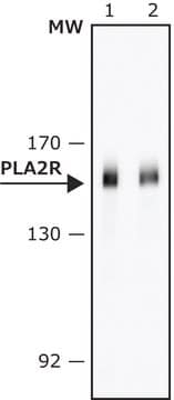
Western Blotting Analysis: 0.5 µg/mL from a representative lot detected PLA2R in 10 µg of human kidney tissue lysate.
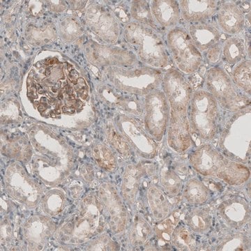
Immunohistochemistry Analysis: A 1:250 dilution from a representative lot detected PLA2R in human kidney tissue.

Quality

Evaluated by Western Blotting in human lung tissue lysate.

Western Blotting Analysis: 0.5 µg/mL of this antibody detected PLA2R in 10 µg of human lung tissue lysate.

Target description

~180 kDa observed. Target band(s) appears larger than the calculated molecular weights (166.6 kDa/isoform 1 and 150.8 kDa/isoform 2) due to posttranslational glycosylation. Uncharacterized band(s) may appear in some lysates.

Physical form

Format: Purified

Other Notes

Concentration: Please refer to lot specific datasheet.

Not finding the right product?  

Try our Product Selector Tool.

Storage Class Code

12 - Non Combustible Liquids

WGK

WGK 1

Flash Point(F)

Not applicable

Flash Point(C)

Not applicable


Certificates of Analysis (COA)

Search for Certificates of Analysis (COA) by entering the products Lot/Batch Number. Lot and Batch Numbers can be found on a product’s label following the words ‘Lot’ or ‘Batch’.

Already Own This Product?

Find documentation for the products that you have recently purchased in the Document Library.

Visit the Document Library

Astgik Petrosyan et al.
Nature communications, 10(1), 3656-3656 (2019-08-15)
In this work we model the glomerular filtration barrier, the structure responsible for filtering the blood and preventing the loss of proteins, using human podocytes and glomerular endothelial cells seeded into microfluidic chips. In long-term cultures, cells maintain their morphology
Qi Zhang et al.
JCI insight, 9(4) (2024-01-16)
The deposition of antipodocyte autoantibodies in the glomerular subepithelial space induces primary membranous nephropathy (MN), the leading cause of nephrotic syndrome worldwide. Taking advantage of the glomerulus-on-a-chip system, we modeled human primary MN induced by anti-PLA2R antibodies. Here we show
Shahbaz Khan et al.
Acta neuropathologica communications, 12(1), 39-39 (2024-03-08)
Chordomas are clinically aggressive tumors with a high rate of disease progression despite maximal therapy. Given the limited therapeutic options available, there remains an urgent need for the development of novel therapies to improve clinical outcomes. Cell surface proteins are attractive

Our team of scientists has experience in all areas of research including Life Science, Material Science, Chemical Synthesis, Chromatography, Analytical and many others.

Contact Technical Service