Skip to Content
Merck
All Photos(1)

Key Documents

313M-1

Sigma-Aldrich

Cytokeratin Cocktail (AE1 & AE3) Mouse Monoclonal Antibody

Sign Into View Organizational & Contract Pricing


About This Item

UNSPSC Code:
12352203
NACRES:
NA.41
clone:
AE1 & AE3, monoclonal
application:
IHC (p)
species reactivity:
human
technique(s):
immunohistochemistry (formalin-fixed, paraffin-embedded sections): 1:100-1:500
citations:
4

biological source

mouse

Quality Level

100
500

conjugate

unconjugated

antibody form

culture supernatant

antibody product type

primary antibodies

clone

AE1 & AE3, monoclonal

description

For In Vitro Diagnostic Use in Select Regions (See Chart)

form

buffered aqueous solution

species reactivity

human

packaging

vial of 0.1 mL concentrate (313M-14)
vial of 0.5 mL concentrate (313M-15)
bottle of 1.0 mL predilute (313M-17)
vial of 1.0 mL concentrate (313M-16)
bottle of 7.0 mL predilute (313M-18)

manufacturer/tradename

Cell Marque

technique(s)

immunohistochemistry (formalin-fixed, paraffin-embedded sections): 1:100-1:500

isotype

IgG1κ

control

breast, colon, prostate, skin

shipped in

wet ice

storage temp.

2-8°C

visualization

cytoplasmic

Gene Information

human ... KRT1(3848)

General description

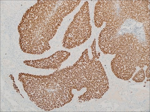
Anti-cytokeratin cocktail (AE1 & AE3) is a broad-spectrum keratin antibody cocktail. It is composed of mouse monoclonal antibody AE1 that recognizes acidic type I keratins and AE3 that reacts with basic type II keratins. Both clones were generated using epidermal keratin as immunogen. This antibody is useful for identifying cells of epithelial origin and detects most carcinomas of different primarysites. This antibody cocktail can cross-react with other intermediate filaments, such as glialfibrillary acidic protein, giving false-positive staining in glial tumors.

Quality


IVD

IVD

IVD

RUO

Linkage

Cytokeratin Cocktail Positive Control Slides, Product No. 313S, are available for immunohistochemistry (formalin-fixed, paraffin-embedded sections).

Physical form

Solution in Tris Buffer, pH 7.3-7.7, with 1% BSA and <0.1% Sodium Azide

Preparation Note

Download the IFU specific to your product lot and formatNote: This requires a keycode which can be found on your packaging or product label.

Other Notes

For Technical Service please contact: 800-665-7284 or email: service@cellmarque.com

Legal Information

Cell Marque is a trademark of Merck KGaA, Darmstadt, Germany

Not finding the right product?  

Try our Product Selector Tool.


Choose from one of the most recent versions:

Certificates of Analysis (COA)

Lot/Batch Number

Don't see the Right Version?

If you require a particular version, you can look up a specific certificate by the Lot or Batch number.

Already Own This Product?

Find documentation for the products that you have recently purchased in the Document Library.

Visit the Document Library

V K Kriho et al.
Virchows Archiv : an international journal of pathology, 431(2), 139-147 (1997-08-01)
Several studies have shown that immunoenzymatic staining of formalin-fixed, paraffin-embedded astrocytomas with keratin antibodies frequently yields positive labelling, but no biochemical evidence of keratin expression in astrocytomas has been reported. We have investigated the presence of keratin in astrocytoma and
Classification of human epithelia and their neoplasms using monoclonal antibodies to keratins: strategies, applications, and limitations.
D Cooper et al.
Laboratory investigation; a journal of technical methods and pathology, 52(3), 243-256 (1985-03-01)
A M Gown et al.
American journal of clinical pathology, 84(4), 413-424 (1985-10-01)
A panel of monoclonal antibodies to human intermediate filament proteins was tested on an unselected series of 246 neoplasms. The antibody panel includes two different anti-cytokeratin antibodies, an anti-vimentin antibody, and an anti-neurofilament antibody (Gown and Vogel, Am J Pathol
H Battifora
The American journal of surgical pathology, 12 Suppl 1, 24-42 (1988-01-01)
The past few years have seen great progress in our knowledge of the biology and the chemistry of intermediate filaments and other filamentous proteins. This has translated into the development of antibodies that, together with new, highly sensitive immunohistochemical methods

Related Content

Diagnostic immunohistochemistry overview highlights its importance in cancer diagnosis and detecting infectious diseases in modern clinical pathology.

Trusted IVDR-ready supplier of high-quality Cell Marque™ antibodies for diagnostic pathology and IHC, ensuring reliability and expert support.

Our team of scientists has experience in all areas of research including Life Science, Material Science, Chemical Synthesis, Chromatography, Analytical and many others.

Contact Technical Service