Skip to Content
Merck
All Photos(1)

Documents

Safety Information

MABS461

Sigma-Aldrich

Anti-Neutrophil Elastase Antibody, clone AHN-10

clone AHN-10, from mouse

Synonym(s):

Neutrophil elastase, Bone marrow serine protease, Elastase-2, Human leukocyte elastase, HLE, Medullasin, PMN elastase

Sign Into View Organizational & Contract Pricing


About This Item

UNSPSC Code:
12352203
eCl@ss:
32160702
NACRES:
NA.41

biological source

mouse

Quality Level

antibody form

purified immunoglobulin

antibody product type

primary antibodies

clone

AHN-10, monoclonal

species reactivity

human

technique(s)

immunocytochemistry: suitable
immunohistochemistry: suitable
immunoprecipitation (IP): suitable
inhibition assay: suitable
radioimmunoassay: suitable

isotype

IgG1κ

NCBI accession no.

UniProt accession no.

shipped in

wet ice

target post-translational modification

unmodified

Gene Information

human ... ELANE(1991)

General description

Neutrophil Elastase, also known as Bone marrow serine protease, Elastase-2, Human leukocyte elastase (HLE), Medullasin or PMN elastase, and encoded by the gene ELANE/ELA2, is a serine proteinase in the same family as chymotrypsin. Neutrophil Elastase has broad substrate specificity and is part of our host defense mechanism against bacteria. Unfortunately passively released or actively secreted elastase from neutrophils has been linked to the pathologic processes of a variety of inflammatory diseases, including idiopathic pulmonary fibrosis, rheumatoid arthritis, adult respiratory distress syndrome, and cystic fibrosis. Neutrophil Elastase is localized to the cytoplasm in granules. Neutrophil Elastase is expressed in neutrophils and macrophages. Defects in Neutrophil Elastase may be associated with cyclic neutropenia and congential neutropenia diseases where neutrophils fail to mature leaving the patient very vunderable to infections.

Specificity

This antibody detects with human Neutrophil Elastase but not other major neutrophil proteases: Cathapsin G, proteinase 3, collagenase, or esterase N (Skubitz, K. M., et al. (1988). J Leukoc Biol. 44(3):158-165.).

Immunogen

Whole cells corresponding to human Neutrophil Elastase.

Application

Detect Elastase using this mouse monoclonal antibody, Anti-Neutrophil Elastase Antibody, clone AHN-10 validated for use in ICC, IHC, IP, Radioimmunoassay & Inhibition.
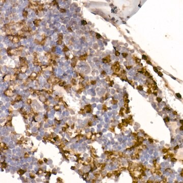
Immunohistochemistry Analysis: A representative lot from an independent laboratory detected Neutrophil Elastase in unirradiated and irradiated human and mouse skin tissues (Rhodes, L. E., et al. (2009). FASEB J. 23(11): 3947-3956.; Nicolaou, A., et al. (2012). Photochem Photobiol Sci. 11(2):371-380.; Rutter, J. J., et al. (2009). 129(12):2861-2867.).

Western Blotting Analysis: A representative lot from an independent laboratory detected Neutrophil Elastase in human pancreatic tissue lysate (Mayerle, J., et al. (2005). Gastroenterology. 129(4):1251-1267.).

Immunoprecipitation Analysis: A representative lot from an independent laboratory immunoprecipitated Neutrophil Elastase in human pancreatic tissue lysate (Mayerle, J., et al. (2005). Gastroenterology. 129(4):1251-1267.).

Immunocytochemistry Analysis: A representative lot from an independent laboratory detected Neutrophil Elastase in human acinar cells (Mayerle, J., et al. (2005). Gastroenterology. 129(4):1251-1267.).

Inhibition Analysis: This assay inhibits the elastinolytic actiity of purified human Neutrophil Elastase (Skubitz, K., et al. (1988). J Leukoc Biol. 44(3):158-165.).

Radioimmunoassay Analysis: A representative lot from an independent laboratory detected Neutrophil Elastase using purified Neutrophil Elastase (Skubitz, K., et al. (1988). J Leukoc Biol. 44(3):158-165.).
Research Category
Cell Structure
Research Sub Category
ECM Proteins

Quality

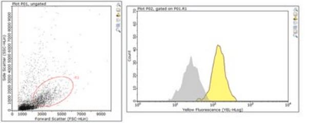
Evaluated by Immunocytochemistry Analysis in peripheral blood mononuclear cells.

Immunocytochemistry Analysis: A 1:250 dilution of this antibody detected Neutrophil Elastase in peripheral blood mononuclear cells.

Target description

28 kDa calculated

Physical form

Format: Purified
Protein G Purified
Purified mouse monoclonal IgG1κ in buffer containing 0.1 M Tris-Glycine (pH 7.4), 150 mM NaCl with 0.05% sodium azide.

Storage and Stability

Stable for 1 year at 2-8°C from date of receipt.

Other Notes

Concentration: Please refer to the Certificate of Analysis for the lot-specific concentration.

Disclaimer

Unless otherwise stated in our catalog or other company documentation accompanying the product(s), our products are intended for research use only and are not to be used for any other purpose, which includes but is not limited to, unauthorized commercial uses, in vitro diagnostic uses, ex vivo or in vivo therapeutic uses or any type of consumption or application to humans or animals.

Not finding the right product?  

Try our Product Selector Tool.

Storage Class Code

12 - Non Combustible Liquids

WGK

WGK 2

Flash Point(F)

Not applicable

Flash Point(C)

Not applicable


Regulatory Listings

Regulatory Listings are mainly provided for chemical products. Only limited information can be provided here for non-chemical products. No entry means none of the components are listed. It is the user’s obligation to ensure the safe and legal use of the product.

JAN Code

MABS461:


Certificates of Analysis (COA)

Search for Certificates of Analysis (COA) by entering the products Lot/Batch Number. Lot and Batch Numbers can be found on a product’s label following the words ‘Lot’ or ‘Batch’.

Already Own This Product?

Find documentation for the products that you have recently purchased in the Document Library.

Visit the Document Library

Maria Tokuyama et al.
The Journal of experimental medicine, 218(7) (2021-05-22)
Neutrophil activation and the formation of neutrophil extracellular traps (NETs) are hallmarks of innate immune activation in systemic lupus erythematosus (SLE). Here we report that the expression of an endogenous retrovirus (ERV) locus ERV-K102, encoding an envelope protein, was significantly
Cristina de Diego et al.
Respiratory research, 25(1), 48-48 (2024-01-20)
Neutrophil extracellular traps (NETs) have repeatedly been related to COVID-19 severity and mortality. However, there is no consensus on their quantification, and there are scarce data on their evolution during the disease. We studied circulating NET markers in patients with

Our team of scientists has experience in all areas of research including Life Science, Material Science, Chemical Synthesis, Chromatography, Analytical and many others.

Contact Technical Service