Skip to Content
Merck
All Photos(1)

Documents

Safety Information

05-510

Sigma-Aldrich

Anti-TCF-1 Antibody, clone 7H3

clone 7H3, Upstate®, from mouse

Sign Into View Organizational & Contract Pricing


About This Item

UNSPSC Code:
12352203
eCl@ss:
32160702
NACRES:
NA.41

biological source

mouse

antibody form

purified immunoglobulin

antibody product type

primary antibodies

clone

7H3, monoclonal

species reactivity

human

manufacturer/tradename

Upstate®

technique(s)

immunocytochemistry: suitable
immunohistochemistry: suitable
western blot: suitable

isotype

IgG

NCBI accession no.

UniProt accession no.

shipped in

wet ice

target post-translational modification

unmodified

Gene Information

human ... HNF1A(6927)

General description

TCF 1 (T cell factor 1) is a member of the TCF/LEF (lymphoid enhancer factor) family of transcription factors. Members of this family along with b catenin, are involved in mediating Wnt signaling by transactivating downstream genes. TCF 1 is closely related to LEF-1, both of which are transcription factors expressed in mice during T cell differentiation. They regulate the T cell receptor a enhancer in transfections assays. TCF 1 becomes transcriptionally active through interaction with b catenin. Also, numerous splice variants of TCF 1 have been identified. Overexpression of TCF-1-specific splice forms appears to correlate with the metastatic behavior of colorectal cancer cells and with overproduction of lymphoid tyrosine protein kinase p56(lck).

Specificity

Recognizes multiple splice isoforms of TCF-1.

Immunogen

Maltose binding protein (MBP) fusion protein corresponding to residues 102-269 of human TCF-1A splice variant. The epitope maps to a region just N-terminal to the High Mobility Group (HMG) box

Application

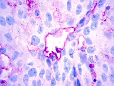
Detect TCF-1 with Anti-TCF-1 Antibody, clone 7H3 (Mouse Monoclonal Antibody), that has been shown to work in ICC, IHC & WB.
Research Category
Epigenetics & Nuclear Function
Research Sub Category
Transcription Factors

Quality

routinely evaluated by immunoblot on Jurkat RIPA cell lysate

Target description

26 kDa, 32 kDa, 38 kDa, 42 kDa, and 48 kDa

Linkage

Replaces: 04-1159

Physical form

Format: Purified
Protein G Purified
Protein G Purified immunoglobulin in Immunoaffinity Purified immunoglobulin in 0.2M Tris-glycine, pH 7.5, 0.15M NaCl and 0.05% sodium azide

Storage and Stability

Maintain for 2 years at -20°C from date of shipment. Aliquot to avoid repeated freezing and thawing. For maximum recovery of product, centrifuge the original vial after thawing and prior to removing the cap.

Analysis Note

Control
Positive Antigen Control: Catalog #12-303, Jurkat cell lysate.

Other Notes

Concentration: Please refer to the Certificate of Analysis for the lot-specific concentration.

Legal Information

UPSTATE is a registered trademark of Merck KGaA, Darmstadt, Germany

Disclaimer

Unless otherwise stated in our catalog or other company documentation accompanying the product(s), our products are intended for research use only and are not to be used for any other purpose, which includes but is not limited to, unauthorized commercial uses, in vitro diagnostic uses, ex vivo or in vivo therapeutic uses or any type of consumption or application to humans or animals.

Not finding the right product?  

Try our Product Selector Tool.

Storage Class Code

12 - Non Combustible Liquids

WGK

WGK 1

Flash Point(F)

Not applicable

Flash Point(C)

Not applicable


Regulatory Listings

Regulatory Listings are mainly provided for chemical products. Only limited information can be provided here for non-chemical products. No entry means none of the components are listed. It is the user’s obligation to ensure the safe and legal use of the product.

JAN Code

05-510:


Certificates of Analysis (COA)

Search for Certificates of Analysis (COA) by entering the products Lot/Batch Number. Lot and Batch Numbers can be found on a product’s label following the words ‘Lot’ or ‘Batch’.

Already Own This Product?

Find documentation for the products that you have recently purchased in the Document Library.

Visit the Document Library

Haiyan Li et al.
American journal of physiology. Cell physiology, 299(2), C422-C430 (2010-07-29)
In our previous studies, roles of gap junction and vascular endothelial growth factor in the cross-talking of human bone marrow stromal cells (HBMSCs) and human umbilical vein endothelial cells (HUVECs) have been extensively studied. The present study focused on the
M Van de Wetering et al.
Molecular and cellular biology, 16(3), 745-752 (1996-03-01)
Previously, we reported the isolation of cDNA clones representing four alternative splice forms of TCF-1, a T-cell-specific transcription factor. In the present study, Western blotting (immunoblotting) yielded a multitude of TCF-1 proteins ranging from 25-55 kDa, a pattern not simply
Weichun He et al.
Journal of the American Society of Nephrology : JASN, 23(2), 294-304 (2011-11-19)
A variety of chronic kidney diseases exhibit reactivation of Wnt/β-catenin signaling. In some tissues, β-catenin transcriptionally regulates matrix metalloproteinase-7 (MMP-7), but the association between MMP-7 and Wnt/β-catenin signaling in chronic kidney disease is unknown. Here, in mouse models of both
Weichun He et al.
The Journal of biological chemistry, 285(32), 24665-24675 (2010-06-04)
Plasminogen activator inhibitor-1 (PAI-1) is a multifunctional glycoprotein that plays a critical role in the pathogenesis of chronic kidney and cardiovascular diseases. Although transforming growth factor (TGF)-beta1 is a known inducer of PAI-1, how it controls PAI-1 expression remains enigmatic.

Our team of scientists has experience in all areas of research including Life Science, Material Science, Chemical Synthesis, Chromatography, Analytical and many others.

Contact Technical Service