MAB329-C
Anti-Synaptophysin Antibody, clone SP15 (Ascites Free)
clone SP15, from mouse
Synonym(s):
Major synaptic vesicle protein p38, Synaptophysin
About This Item
Recommended Products
biological source
mouse
Quality Level
antibody form
purified antibody
antibody product type
primary antibodies
clone
SP15, monoclonal
species reactivity
human, monkey, feline, rat, mouse
technique(s)
ELISA: suitable
immunocytochemistry: suitable
immunohistochemistry: suitable
western blot: suitable
isotype
IgMκ
NCBI accession no.
UniProt accession no.
target post-translational modification
unmodified
Gene Information
human ... SYP(6855)
General description
Immunogen
Application
Neuroscience
Developmental Neuroscience
Western Blotting Analysis: A representative lot detected synaptophysin in rat hippocampal tissue extracts (Lin, D., et al. (2012). Behav. Brain Res. 228(2):319-327).
Western Blotting Analysis: Representative lots detected synaptophysin in mouse brain synaptosomes preparations (Shim, S.Y., et al. (2008). J. Neurosci. 28(14):3604-3614).
Western Blotting Analysis: A representative lot detected synaptophysin in the large dense-core vesicles-containing fractions obtained by sucrose gradient centrifugation of rat brain median eminence (ME) neuroterminals preparations (Yin, W., et al. (2007). Exp. Biol. Med. (Maywood). 232(5):662-673).
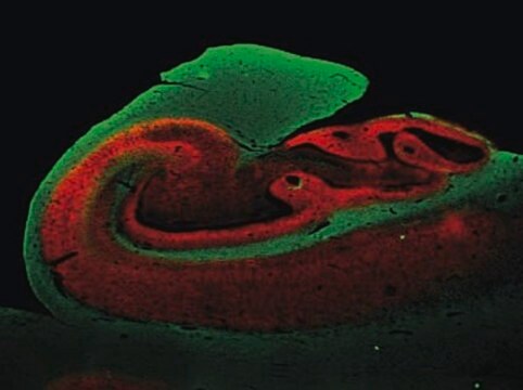
Immunohistochemistry Analysis: A representative lot detected a stronger synaptophysin immunoreactivity in the inner molecular layer of frozen dentate gyrus sections from a 3-week old monkey, while a stronger immunoreactivity was seen in the outer molecular layer of dentate gyrus sections from a 13-year old monkey (Lavenex, P., et al. (2007). Dev. Neurosci. 29(1-2):179–192).
Immunohistochemistry Analysis: A representative lot immunostained nerve terminals at palisade ending using cat extraocular muscle (EOM) whole mount sections (Konakci, K.Z., et al. (2005). Invest. Ophthalmol. Vis. Sci. 46(1):155-165).
Immunohistochemistry Analysis: A representative lot detected synaptophysin immunoreactivity in human brain tissue sections (Honer, W.G., et al. (1993). Brain Res. 609(1-2):9-20).
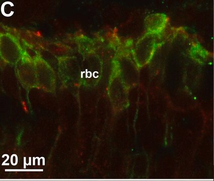
Immunocytochemistry Analysis: A representative lot detected punctate synatophysin immunoreactivity among cultured primary cerebrocortical neurons from rat embryos (Bragina, L., et al. (2006). J. Neurochem. 99(1):134-141.).
ELISA Analysis: Representative lots were employed as the capture antibody for the detection of synaptophysin in human temporal cortex, frontal cortex, and cerebellar cortex extracts by sandwich ELISA (Klucken, J., et al. (2006). Acta Neuropathol. 111(2):101-108; Fukumoto, H., et al. (2002). Arch. Neurol. 59(9):1381-1389).
Quality
Western Blotting Analysis: 1.0 µg/mL of this antibody detected Synaptophysin in 10 µg of rat brain tissue lysate.
Target description
Physical form
Storage and Stability
Other Notes
Disclaimer
Not finding the right product?
Try our Product Selector Tool.
recommended
Storage Class Code
12 - Non Combustible Liquids
WGK
WGK 2
Flash Point(F)
Not applicable
Flash Point(C)
Not applicable
Certificates of Analysis (COA)
Search for Certificates of Analysis (COA) by entering the products Lot/Batch Number. Lot and Batch Numbers can be found on a product’s label following the words ‘Lot’ or ‘Batch’.
Already Own This Product?
Find documentation for the products that you have recently purchased in the Document Library.
Our team of scientists has experience in all areas of research including Life Science, Material Science, Chemical Synthesis, Chromatography, Analytical and many others.
Contact Technical Service


