Skip to Content
Merck
All Photos(3)

Key Documents

05-784R

Sigma-Aldrich

Anti-IRS1 Antibody, clone 58-10C-31, rabbit monoclonal

clone 58-10C-31, from rabbit

Synonym(s):

insulin receptor substrate 1

Sign Into View Organizational & Contract Pricing


About This Item

UNSPSC Code:
12352203
eCl@ss:
32160702
NACRES:
NA.41

biological source

rabbit

Quality Level

antibody form

purified immunoglobulin

antibody product type

primary antibodies

clone

58-10C-31, monoclonal

species reactivity

human, rat, mouse

technique(s)

immunoprecipitation (IP): suitable
western blot: suitable

isotype

IgG

NCBI accession no.

UniProt accession no.

shipped in

dry ice

target post-translational modification

unmodified

Gene Information

human ... IRS1(3667)

General description

IRS1 (Insulin Receptor Substrate 1) transmits insulin signals via metabolic and mitogenic pathways. It is heavily phosphorylated on both serine and tyrosine residues. These phosphorylated tyrosines enable IRS to act as a docking protein that binds SH2 domains of such proteins as PI3 Kinase (phosphatidylinositol 3-kinase) and GRB2, resulting in activation. Over expression and phosphorylation of serine is associated with insulin resistance and breast cancer.

Specificity

Antibody recognizes the C-terminus of IRS1.

Immunogen

Epitope: C-terminus
KLH- conjugated - linear peptide corresponding to the C-terminus of rat IRS1.

Application

Detect IRS1 using this Anti-IRS1 Antibody, clone 58-10C-31 validated for use in WB & IP.
Research Category
Metabolism
Research Sub Category
Insulin/Energy Signaling
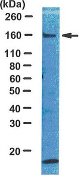
Western Blot (SNAP ID) Analysis: 0.1 µg/mL from a previous lot detected IRS-1 on 10 µg of 3T3/A31, 3T3/L1, L6 and MCF7 cell lysates.

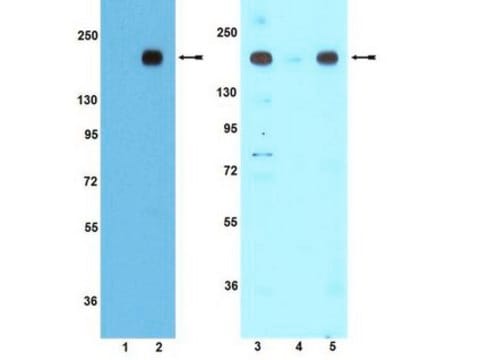
Immunoprecipitation Analysis: 1 µg from a previous lot immunoprecipitated IRS-1 from 100 µg of MCF7 cell lysate. Arrow indicates IRS-1 (~160 kDa). There is cross reactivity of the detection antibody with the heavy chaing of Rabbit IgG as shown at ~50 kDa.

Quality

Evaluated by Western Blot in 3T3/A31, 3T3/L1, L6 or MCF7 cell lysates.

Western Blot Analysis: 0.1 µg/mL of this antibody detected IRS-1 on 10 µg of 3T3/A31, 3T3/L1, L6, or MCF7 cell lysates.

Target description

~ 160 kDa

Physical form

Format: Purified
Protein A purified
Purified Rabbit Monoclonal IgG Supernatant in buffer containing 0.1 M Tris-Glycine (pH 7.4, 150 mM NaCl) with 0.05% sodium azide and 40% glycerol.

Storage and Stability

Stable for 1 year at -20°C from date of receipt.
Handling Recommendations: Upon receipt and prior to removing the cap, centrifuge the vial and gently mix the solution. Aliquot into microcentrifuge tubes and store at -20°C. Avoid repeated freeze/thaw cycles, which may damage IgG and affect product performance.
Note: Variability in freezer temperatures below -20°C may cause glycerol containing solutions to become frozen during storage.

Analysis Note

Control
3T3/A31, 3T3/L1, L6, or MCF7 cell lysates

Other Notes

Concentration: Please refer to the Certificate of Analysis for the lot-specific concentration.

Disclaimer

Unless otherwise stated in our catalog or other company documentation accompanying the product(s), our products are intended for research use only and are not to be used for any other purpose, which includes but is not limited to, unauthorized commercial uses, in vitro diagnostic uses, ex vivo or in vivo therapeutic uses or any type of consumption or application to humans or animals.

Not finding the right product?  

Try our Product Selector Tool.

Storage Class Code

12 - Non Combustible Liquids

WGK

WGK 1

Flash Point(F)

Not applicable

Flash Point(C)

Not applicable


Certificates of Analysis (COA)

Search for Certificates of Analysis (COA) by entering the products Lot/Batch Number. Lot and Batch Numbers can be found on a product’s label following the words ‘Lot’ or ‘Batch’.

Already Own This Product?

Find documentation for the products that you have recently purchased in the Document Library.

Visit the Document Library

Hua Yan et al.
Molecular medicine reports, 15(1), 180-186 (2016-12-03)
Nonalcoholic fatty liver disease (NAFLD) is a common chronic liver disease, the pathological process of which is complex. Activation of the c‑Jun N‑terminal kinase (JNK) signaling pathway is associated with the mechanism underlying obesity-induced insulin resistance. Furthermore, the JNK signaling
Rongya Tao et al.
Nature medicine, 24(7), 1058-1069 (2018-06-06)
Unsuppressed hepatic glucose production (HGP) contributes substantially to glucose intolerance and diabetes, which can be modeled by the genetic inactivation of hepatic insulin receptor substrate 1 (Irs1) and Irs2 (LDKO mice). We previously showed that glucose intolerance in LDKO mice
Kyle D Copps et al.
The Journal of biological chemistry, 291(16), 8602-8617 (2016-02-06)
Constitutive activation of the mammalian target of rapamycin complex 1 and S6 kinase (mTORC1→ S6K) attenuates insulin-stimulated Akt activity in certain tumors in part through "feedback" phosphorylation of the upstream insulin receptor substrate 1 (IRS1). However, the significance of this
Pei-Wen Cheng et al.
Diabetes, 64(10), 3413-3424 (2015-04-18)
Aberrant Wnt signaling appears to play an important role in the onset of diabetes. Moreover, the insulin signaling pathway is defective in the nucleus tractus solitarii (NTS) of spontaneously hypertensive rats (SHRs) and fructose-fed rats. Nevertheless, the relationships between Wnt
Himani Kukreti et al.
Journal of cellular biochemistry, 121(5-6), 3070-3089 (2020-02-15)
Aging skeletal muscle shows perturbations in metabolic functions. MicroRNAs have been shown to play a critical role in aging and metabolic functions of skeletal muscle. MicroRNA-34a (miR-34a) is implicated in the brain and cardiac aging, however, its role in aging

Our team of scientists has experience in all areas of research including Life Science, Material Science, Chemical Synthesis, Chromatography, Analytical and many others.

Contact Technical Service