Skip to Content
Merck
All Photos(2)

Key Documents

HPA011138

Sigma-Aldrich

ANTI-APPL1 antibody produced in rabbit

Prestige Antibodies® Powered by Atlas Antibodies, affinity isolated antibody, buffered aqueous glycerol solution

Synonym(s):

Anti-APPL, Anti-Adapter protein containing PH domain, PTB domain and leucine zipper motif 1, Anti-DCC-interacting protein 13-alpha, Anti-Dip13-alpha

Sign Into View Organizational & Contract Pricing


About This Item

UNSPSC Code:
12352203
Human Protein Atlas Number:
NACRES:
NA.41
conjugate:
unconjugated
application:
IHC
clone:
polyclonal
species reactivity:
human
citations:
4
technique(s):
immunohistochemistry: 1:500- 1:1000

biological source

rabbit

Quality Level

conjugate

unconjugated

antibody form

affinity isolated antibody

antibody product type

primary antibodies

clone

polyclonal

product line

Prestige Antibodies® Powered by Atlas Antibodies

form

buffered aqueous glycerol solution

species reactivity

human

technique(s)

immunohistochemistry: 1:500- 1:1000

immunogen sequence

VTRLTFPLPCVVLYATHQENKRLFGFVLRTSSGRSESNLSSVCYIFESNNEGEKICDSVGLAKQIALHAELDRRASEKQKEIERVKEKQQKELNKQKQIEKDLEEQ

UniProt accession no.

shipped in

wet ice

storage temp.

−20°C

target post-translational modification

unmodified

Gene Information

human ... APPL1(26060)

General description

APPL1 (adaptor protein, phosphotyrosine interaction, PH domain and leucine zipper containing 1) is an endosomal adaptor protein which is made of 709 amino acids. It has a PTB (phosphotyrosine binding) domain at its C-terminal, Bin-Amphiphysin-Rvs (BAR) domain at the N-terminal, and an intervening pleckstrin homology (PH) domain. This gene is localized to the human chromosomal region 3p14.3-p21.1. It is abundantly expressed in heart, pancreas, ovary and skeletal muscle.

Immunogen

DCC-interacting protein 13-alpha recombinant protein epitope signature tag (PrEST)

Application

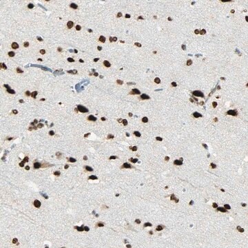
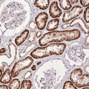
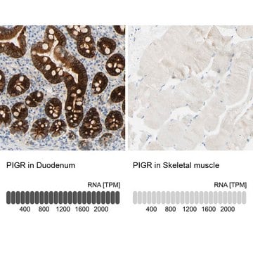
All Prestige Antibodies Powered by Atlas Antibodies are developed and validated by the Human Protein Atlas (HPA) project and as a result, are supported by the most extensive characterization in the industry.

The Human Protein Atlas project can be subdivided into three efforts: Human Tissue Atlas, Cancer Atlas, and Human Cell Atlas. The antibodies that have been generated in support of the Tissue and Cancer Atlas projects have been tested by immunohistochemistry against hundreds of normal and disease tissues and through the recent efforts of the Human Cell Atlas project, many have been characterized by immunofluorescence to map the human proteome not only at the tissue level but now at the subcellular level. These images and the collection of this vast data set can be viewed on the Human Protein Atlas (HPA) site by clicking on the Image Gallery link. We also provide Prestige Antibodies® protocols and other useful information.

Biochem/physiol Actions

APPL1 (adaptor protein, phosphotyrosine interaction, PH domain and leucine zipper containing 1) is an adaptor protein for AKT2 (v-akt murine thymoma viral oncogene homolog 2) and p110α, and anchors them to cytoplasm. This speeds the recruitment of AKT2 and p110α to cell membrane during cell division. Upon stimulation by epidermal growth factor (EGF) or oxidative stress, it moves from cell membrane to nucleus. It is therefore, involved in regulating transcription and chromatin modelling, by interacting with nucleosome remodeling and histone deacetylase multiprotein complex NuRD/MeCP1. APPL1 suppresses AKT by preventing Src-mediated phosphorylation of tyrosine residues. It thus, inhibits AKT mediated cell migration and adhesion. The BAR domain is responsible for sensing and inducing membrane curvature, and the PH domains interacts with phosphoinositols. It contributes to the development of synapses and dendritic spines in hippocampal neurons, and might play a role in memory impairment in Alzheimer′s disease (AD).

Features and Benefits

Prestige Antibodies® are highly characterized and extensively validated antibodies with the added benefit of all available characterization data for each target being accessible via the Human Protein Atlas portal linked just below the product name at the top of this page. The uniqueness and low cross-reactivity of the Prestige Antibodies® to other proteins are due to a thorough selection of antigen regions, affinity purification, and stringent selection. Prestige antigen controls are available for every corresponding Prestige Antibody and can be found in the linkage section.

Every Prestige Antibody is tested in the following ways:
  • IHC tissue array of 44 normal human tissues and 20 of the most common cancer type tissues.
  • Protein array of 364 human recombinant protein fragments.

Linkage

Corresponding Antigen APREST72486

Physical form

Solution in phosphate-buffered saline, pH 7.2, containing 40% glycerol and 0.02% sodium azide

Legal Information

Prestige Antibodies is a registered trademark of Merck KGaA, Darmstadt, Germany

Not finding the right product?  

Try our Product Selector Tool.

Storage Class Code

10 - Combustible liquids

WGK

WGK 1

Flash Point(F)

Not applicable

Flash Point(C)

Not applicable

Personal Protective Equipment

dust mask type N95 (US), Eyeshields, Gloves

Choose from one of the most recent versions:

Certificates of Analysis (COA)

Lot/Batch Number

Don't see the Right Version?

If you require a particular version, you can look up a specific certificate by the Lot or Batch number.

Already Own This Product?

Find documentation for the products that you have recently purchased in the Document Library.

Visit the Document Library

Akari Ogawa et al.
Brain research, 1494, 118-124 (2012-12-19)
Adaptor protein containing a PH domain, PTB domain and leucine zipper motif (APPL1) is emerging as a critical regulator of various cellular processes in non-neuronal cells as well as in neurons where it localizes to dendritic spines and synapses. It
Y Mitsuuchi et al.
Oncogene, 18(35), 4891-4898 (1999-09-22)
AKT2 is a serine/threonine kinase implicated in human ovarian and pancreatic cancers. AKT2 is activated by a variety of growth factors and insulin via phosphatidylinositol 3-kinase (PI3K). However, its normal cellular role is not well understood. To gain insight into
Marta Miaczynska et al.
Cell, 116(3), 445-456 (2004-03-16)
Signals generated in response to extracellular stimuli at the plasma membrane are transmitted through cytoplasmic transduction cascades to the nucleus. We report the identification of a pathway directly linking the small GTPase Rab5, a key regulator of endocytosis, to signal
Joshua A Broussard et al.
Molecular biology of the cell, 23(8), 1486-1499 (2012-03-02)
Cell migration is a complex process that requires the integration of signaling events that occur in distinct locations within the cell. Adaptor proteins, which can localize to different subcellular compartments, where they bring together key signaling proteins, are emerging as

Our team of scientists has experience in all areas of research including Life Science, Material Science, Chemical Synthesis, Chromatography, Analytical and many others.

Contact Technical Service