General description
Microtubule-associated protein tau (UniProt: P10636; also known as Neurofibrillary tangle protein, Paired helical filament-tau, PHF-tau) is encoded by the MAPT (also known as MAPTL, MTBT1, TAU) gene (Gene ID: 4137) in human. Microtubule-associated protein tau is shown to be expressed in neurons. It is mostly found in axons, in the cytosol, and in association with plasma membrane components. Nine isoforms of Tau have been described that are generated by alternative splicing. Tau proteins promote microtubule assembly and stability and may also be involved in the establishment and maintenance of neuronal polarity. The C-terminus binds axonal microtubules while the N-terminus binds neural plasma membrane components, thus serving as a linker protein between both. The short isoforms are reported to allow plasticity of the cytoskeleton whereas the longer isoforms may preferentially play a role in its stabilization. In Alzheimer disease (AD), the neuronal cytoskeleton in the brain is progressively disrupted and replaced by tangles of paired helical filaments (PHF) and straight filaments, mainly composed of hyperphosphorylated forms of Tau. In AD brain Tau accumulates in both the somatodendritic and axonal domains of neurons and it also accumulates in the soma as neurofibrillary tangles (NFTs). Tau oligomers are reported to be neurotoxic when applied extracellularly to cultured neuronal cells and can induce neurodegeneration and synaptic and mitochondrial dysfunction in vivo. It has been reported that most of the C-terminus of Tau (aa 243-406) in PHFs can be cleaved by trypsin, however the TauC4 region is highly resistant to trypsin action. (Ref.: Taniguchi Watanabe, S., et al. (2016). Acta Neuropathol. 131(2); 267-280; Ando K., et al. (2010). Biochem. Soc. Trans. 38(4); 1001-1005).
Specificity
This rabbit polyclonal antibody specifically detects TauC4 region in human Tau protein.
Immunogen
Epitope: unknown
KLH-conjugated linear peptide corresponding to 16 amino acids from the C-terminal half of human Microtubule-associated protein tau, isoform 8 (Tau-F).
Application
Anti-Tau (TauC4, 354-369), Cat. No. ABN2178, is a highly specific rabbit polyclonal antibody that targets TauC4 region of Tau and has been tested for use in Electron Microscopy and Western Blotting.
Research Category
Neuroscience
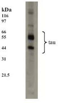
Western Blotting Analysis: A representative lot detected Tau (TauC4, 354-369) in Western Blotting applications (Fitzpatrick, A.W.P., et. al. (2017) Nature. 547(7662):185-190; Taniguchi-Watanabe, S., et. al. (2016). Acta Neuropathol. 131(2):267-280).
Electron Microscopy Analysis: A representative lot detected Tau (TauC4, 354-369) in Immunogold negative-stain Electron Microscopy applications (Fitzpatrick, A.W.P., et. al. (2017) Nature. 547(7662):185-190).
Quality
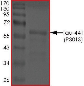
Evaluated by Western Blotting in Recombinant full-length 4-repeat (4R) tau (human 4R1N).
Western Blotting Analysis: A 1:2,000 dilution of this antibody detected TauC4 region in full length recombinant human tau (human 4R1N).
Target description
~55 kDa observed; 45.85 kDa calculated.
Physical form
Rabbit polyclonal antiserum with 0.1% sodium azide.
Unpurified
Storage and Stability
Stable for 1 year at -20°C from date of receipt. Handling Recommendations: Upon receipt and prior to removing the cap, centrifuge the vial and gently mix the solution. Aliquot into microcentrifuge tubes and store at -20°C. Avoid repeated freeze/thaw cycles, which may damage IgG and affect product performance.
Other Notes
Concentration: Please refer to lot specific datasheet.
Disclaimer
Unless otherwise stated in our catalog or other company documentation accompanying the product(s), our products are intended for research use only and are not to be used for any other purpose, which includes but is not limited to, unauthorized commercial uses, in vitro diagnostic uses, ex vivo or in vivo therapeutic uses or any type of consumption or application to humans or animals.


