Skip to Content
Merck
All Photos(6)

Key Documents

P7372

Sigma-Aldrich

Anti-Protein Disulfide Isomerase (MD-12) antibody produced in rabbit

enhanced validation

affinity isolated antibody, buffered aqueous solution

Synonym(s):

Anti-Erp58, Anti-PDI

Sign Into View Organizational & Contract Pricing


About This Item

MDL number:
UNSPSC Code:
12352203
NACRES:
NA.41

biological source

rabbit

Quality Level

conjugate

unconjugated

antibody form

affinity isolated antibody

antibody product type

primary antibodies

clone

polyclonal

form

buffered aqueous solution

mol wt

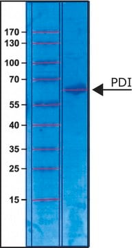
antigen 57 kDa

species reactivity

human, bovine, rat, mouse

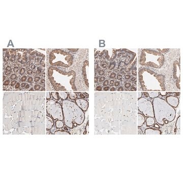
enhanced validation

independent
Learn more about Antibody Enhanced Validation

technique(s)

immunoprecipitation (IP): 2-5 μg using RIPA lysate (500 μg) of rat NRK cells
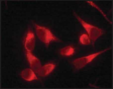
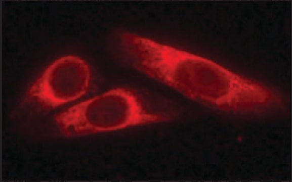
indirect immunofluorescence: 2-5 μg/mL using human HeLa cells
western blot (chemiluminescent): 0.1-0.2 μg/mL using whole extract of mouse NIH3T3 cells

UniProt accession no.

shipped in

dry ice

storage temp.

−20°C

target post-translational modification

unmodified

Gene Information

human ... P4HB(5034)
mouse ... P4hb(18453)
rat ... P4hb(25506)

General description

Anti-Protein Disulfide Isomerase (PDI) (MD-12) is developed in rabbit using as immunogen a synthetic peptide corresponding to amino acid residues of human PDI with N-terminal added cysteine, conjugated to KLH. Protein disulfide isomerase (PDI, Erp58) is an abundant multifunctional, soluble enzyme (E.C. 5.3.4.1) that resides in the lumen of the endoplasmic reticulum of eukaryotic cells. The mammalian PDI family comprises several highly divergent proteins that contain one or more thioredoxin like structural domains. PDI consists of four tandem domains, two of which contain a catalytic site for S-S bond formation. PDI has an N-terminal ER signal and C-terminal ER retention KDEL signal sequences. PDI may also be expressed in other cellular localizations such as the cell surface, cytosol, and nucleus. PDI was found on the cell surface of several cell types including platelets, lymphoid cells, pancreatic exocrine cells, retinal cells, thyroid cells, and hepatocytes.

Immunogen

synthetic peptide corresponding to amino acid residues 495-506 of human protein disulfide isomerase.

Application

Anti-Protein Disulfide Isomerase (MD-12) antibody produced in rabbit has been used in:
  • immunoblotting
  • immunoprecipitation
  • immunofluorescence

Biochem/physiol Actions

Protein disulfide isomerase (PDI) catalyses the formation and rearrangements of both intrachain and interchain disulfide bonds in secreted proteins. PDI also serves as a molecular chaperone that can suppress protein aggregation, or as an anti-chaperone that mediates aggregate formation. PDI respectively participates in the hydroxylation of prolines in procollagen during collagen synthesis and in the transfer of neutral lipid onto nascent lipoprotein particles. PDI has calcium-dependent transglutaminase activity, which catalyses the formation of isopeptide bonds.

Physical form

Solution in 0.01 M phosphate buffered saline containing 1% bovine serum abumin and 15 mM sodium azide.

Disclaimer

Unless otherwise stated in our catalog or other company documentation accompanying the product(s), our products are intended for research use only and are not to be used for any other purpose, which includes but is not limited to, unauthorized commercial uses, in vitro diagnostic uses, ex vivo or in vivo therapeutic uses or any type of consumption or application to humans or animals.

Not finding the right product?  

Try our Product Selector Tool.

Storage Class Code

10 - Combustible liquids

WGK

nwg

Flash Point(F)

Not applicable

Flash Point(C)

Not applicable


Certificates of Analysis (COA)

Search for Certificates of Analysis (COA) by entering the products Lot/Batch Number. Lot and Batch Numbers can be found on a product’s label following the words ‘Lot’ or ‘Batch’.

Already Own This Product?

Find documentation for the products that you have recently purchased in the Document Library.

Visit the Document Library

Hongge Wang et al.
Molecular and cellular biology, 42(6), e0052221-e0052221 (2022-05-03)
Ferroptosis is a form of regulated cell death resulting predominantly from catastrophic accumulation of lipid reactive oxygen species (ROS). While the antioxidant systems that counter ferroptosis have been well characterized, the mechanism underlying ferroptosis-associated accumulation of lipid ROS remains unclear.
Protein disulfide isomerase. A multifunctional protein resident in the lumen of the endoplasmic reticulum.
Noiva R and Lennarz WJ
The Journal of Biological Chemistry, 267(6), 3553-3556 (1992)
Ming-Jie Hou et al.
Acta biochimica et biophysica Sinica, 55(5), 853-865 (2023-05-30)
Ferroptosis is a new form of nonapoptotic cell death closely associated with glutathione (GSH) peroxidase 4 inhibition and/or GSH depletion, resulting in the accumulation of cellular iron and lipid peroxides. The exact mechanism by which GSH depletion causes the accumulation
Dual targeting of the protein disulfide isomerase RB60 to the chloroplast and the endoplasmic reticulum
Levitan A, et al.
Proceedings of the National Academy of Sciences of the USA, 102(17), 6225-6230 (2005)
Xinmiao Fu et al.
The Biochemical journal, 447(1), 115-123 (2012-07-04)
E(2) (17β-oestradiol), a female sex hormone, has important biological functions in a woman's body. The pancreas, often considered a non-classical E(2)-targeting organ, is known to be functionally regulated by E(2), but little is known about how oestrogen actions are regulated

Our team of scientists has experience in all areas of research including Life Science, Material Science, Chemical Synthesis, Chromatography, Analytical and many others.

Contact Technical Service