CBL171-I
Anti-Actin, smooth muscle Antibody, clone ASM-1/1A4
clone ASM-1 (1A4), from mouse
Synonym(s):
Actin, aortic smooth muscle, Alpha-actin-2, Cell growth-inhibiting gene 46 protein
About This Item
Recommended Products
biological source
mouse
Quality Level
antibody form
purified immunoglobulin
antibody product type
primary antibodies
clone
ASM-1 (1A4), monoclonal
species reactivity
mouse, chicken, rat, human, bovine
species reactivity (predicted by homology)
equine (based on 100% sequence homology)
technique(s)
ELISA: suitable
immunocytochemistry: suitable
immunofluorescence: suitable
immunohistochemistry: suitable
western blot: suitable
isotype
IgG2aκ
NCBI accession no.
UniProt accession no.
shipped in
wet ice
target post-translational modification
unmodified
Gene Information
human ... ACTA2(59)
General description
Specificity
Immunogen
Application
Cell Structure
Cytoskeletal Signaling
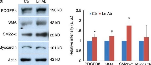
Western Blotting Analysis: A representative lot detected smooth muscle actin in PC-3M-LN4 and PC-3M-LN4-derived human prostate cancer cells (Zvieriev, V., et al. (2005). Biochem. Biophys. Commun. 337(2):498-504).
Western Blotting Analysis: A representative lot detected developing stage-dependent vascular alpha-actin level in chicken heart of various somite stages (Ehler, E., et al. (2004). Dev. Dyn. 229(4):745-755).
Western Blotting Analysis: A representative lot detected aortic actin, but not actin from fibroblasts (beta- and gamma-cytoplasmic), striated muscle (alpha-sarcomeric), or myocardium (alpha-myocardial) in various human, rat, bovine and rabbit tissue and cell lysates (Skalli, O., et al. (1986). J. Cell Biol. 103(6 Pt 2):2787-2796).
Immunohistochemistry Analysis: A representative lot immunostained smooth muscle actin-positive myofibroblasts in paraformaldehyde-fix rat skin wounds sections (Mirastschijski, U., et al. (2010). Wound Repair Regen. 18(2):223-234).
Immunohistochemistry Analysis: A representative lot immunostained activated myofibroblasts in formalin-fixed, paraffin-embedded mouse submandibular salivary gland (Hall, B.E., et al. (2010). Lab. Invest. 90(4):543-555).
Immunohistochemistry Analysis: A representative lot immunostained α-SMA-positive cells in paraffin-embedded rat healing patellar tendon sections (Fu, S.C., et al. (2008). J. Orthop. Res. 26(3):374-383).
Immunocytochemistry Analysis: Representative lots immunostained the mesoderm layer of embryoid bodies formed in vitro from northern white rhinoceros iPSCs, as well as murine iPSCs and mESCs (Ben-Nun, I.F., et al. (2011). Nat. Methods. 8(10):829-831; Gerlach, J.C., et al. (2010). Cells Tissues Organs. 192(1):39-49; Shao, L., et al. (2009). Cell Res. 19(3):296-306).
Immunohistochemistry Analysis: A representative lot immunostained a subsets of cultured rat aortic medial smooth muscle cells (SMCs) and dermal fibroblasts (Skalli, O., et al. (1986). J. Cell Biol. 103(6 Pt 2):2787-2796).
Immunohistochemistry Analysis: A representative lot immunostained cultured rat tendon cells (Fu, S.C., et al. (2008). J. Orthop. Res. 26(3):374-383).
Immunofluorescence Analysis: A representative lot immunostained myofibrils using methanol-fixed, 9-somite stage chicken heart whole-mount preparations. The vascular alpha-actin staining started to disappear from a subset of the myofibrils when using 10-somite stage chicken heart preparations (Ehler, E., et al. (2004). Dev. Dyn. 229(4):745-755).
Immunofluorescence Analysis: A representative lot immunostained stromal vessel SM cells in acetone-fixed benign (leiomyomas) and malignant (leiomyosarcomas & intravascular leiomyomatosis) smooth muscle (SM) neoplasms cryosections (Schürch, W., et al. (1987). Am. J. Pathol. 128(1):91-103).
Immunofluorescence Analysis: A representative lot immunostained chicken gizzard and rat myocardium blood vessels, as well as smooth muscle cells (SMCs) in various acetone-fixed human and rat cryosections, whereas chicken gizzard parenchyrnal cells, rat cardiocytes, aorta endothelial cells and adventitial fibroblasts were negative (Skalli, O., et al. (1986). J. Cell Biol. 103(6 Pt 2):2787-2796).
ELISA Analysis: A representative lot detected aortic actin, but not actin from fibroblasts (beta- and gamma-cytoplasmic), striated muscle (alpha-sarcomeric), or myocardium (alpha-myocardial) by ELISA (Skalli, O., et al. (1986). J. Cell Biol. 103(6 Pt 2):2787-2796).
Quality
Western Blotting Analysis: 0.5 µg/mL of this antibody detected smooth muscle actin in 10 µg of HUVEC lysate.
Target description
Physical form
Storage and Stability
Other Notes
Disclaimer
Not finding the right product?
Try our Product Selector Tool.
Storage Class Code
12 - Non Combustible Liquids
WGK
WGK 1
Flash Point(F)
Not applicable
Flash Point(C)
Not applicable
Certificates of Analysis (COA)
Search for Certificates of Analysis (COA) by entering the products Lot/Batch Number. Lot and Batch Numbers can be found on a product’s label following the words ‘Lot’ or ‘Batch’.
Already Own This Product?
Find documentation for the products that you have recently purchased in the Document Library.
Our team of scientists has experience in all areas of research including Life Science, Material Science, Chemical Synthesis, Chromatography, Analytical and many others.
Contact Technical Service


