Skip to Content
Merck
All Photos(4)

Documents

05-814

Sigma-Aldrich

Anti-HDAC2 Antibody, clone 3F3

clone 3F3, Upstate®, from mouse

Synonym(s):

YY1-associated factor 1, histone deacetylase 2, transcriptional regulator homolog RPD3

Sign Into View Organizational & Contract Pricing


About This Item

UNSPSC Code:
12352203
eCl@ss:
32160702
NACRES:
NA.41

biological source

mouse

Quality Level

antibody form

purified immunoglobulin

antibody product type

primary antibodies

clone

3F3, monoclonal

species reactivity

bovine, human, hamster, rat, mouse

packaging

antibody small pack of 25 μg

manufacturer/tradename

Upstate®

technique(s)

immunocytochemistry: suitable
immunoprecipitation (IP): suitable
western blot: suitable

isotype

IgG1

NCBI accession no.

UniProt accession no.

shipped in

dry ice

target post-translational modification

unmodified

Gene Information

human ... HDAC2(3066)

General description

Histone deacetylase 2 (HDAC2), or transcriptional regulator homolog RPD3 L1, is highly homologous to the yeast transcription factor RPD3 (reduced potassium dependency 3) gene. As in yeast, human HDA2 is likely to be involved in regulating chromatin structure during transcription. It has been implicated to associate with YY1, a mammalian zinc-finger transcription factor, which negatively regulates transcription by tethering RPD3 to DNA as a cofactor. This process is highly conserved from yeast to human.

Specificity

Predicted to cross-react with rat based on sequence homology.
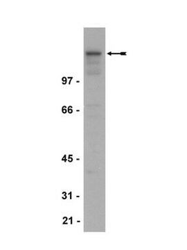
Recognizes HDAC2, MW ~60 kDa.

Immunogen

KLH-conjugated, synthetic peptide (CEKTDTKGTKSEQLSNP) corresponding to amino acids 473-488 of human HDAC2 (Accession # NP_001518) containing a N-terminal cysteine

Application

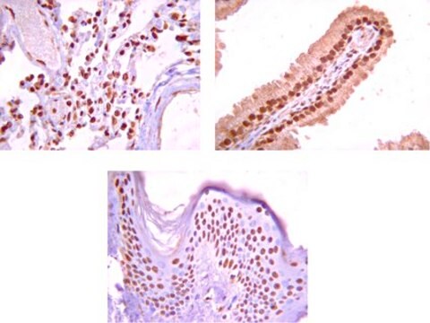
Anti-HDAC2 Antibody, clone 3F3 is a Mouse Monoclonal Antibody for detection of HDAC2 also known as YY1-associated factor 1, histone deacetylase 2 & has been validated in ICC, IP & WB.
Immunocytochemistry:
This antibody has been reported by an independent laboratory to show positive immunostaining for HDAC2 in NIH/3T3 and U2OS cells fixed with paraformaldehyde.

Immunoprecipitation and HDAC Assay:
3 μL of a previous lot was used to immunoprecipitate HDAC activity from HeLa nuclear extract, which was then measured using the HDAC Assay Kit (Fluorometric Detection) (Catalog # 17-356). An independent laboratory has also demonstrated positive immunoprecipitation /HDAC assay results using NIH/3T3 cells.
Research Category
Epigenetics & Nuclear Function
Research Sub Category
Histones

Quality

Evaluated by western blot on nuclear extracts from HeLa cells.

Western Blot Analysis:
0.5-2 μg/mL of this antibody detected HDAC2 in nuclear extracts from HeLa cells.

Target description

~60 kDa

Linkage

Replaces: 04-229

Physical form

Format: Purified
Protein G Purified
Purified mouse monoclonal IgG in buffer containing 70% storage buffer (0.1 M Tris-glycine, pH 7.4, 0.15 M NaCl, 0.05% sodium azide) and 30% glycerol. Store at -20°C.

Storage and Stability

Stable for 1 year at -20ºC from date of receipt.

Analysis Note

Control
Positive Antigen Control: Catalog #12-309, Hela cell nuclear extract. Add an equal volume of Laemmli reducing sample buffer to 10 μL of extract and boil for 5 minutes to reduce the preparation. Load 20 μg of reduced extract per lane for minigels.

Other Notes

Concentration: Please refer to the Certificate of Analysis for the lot-specific concentration.

Legal Information

UPSTATE is a registered trademark of Merck KGaA, Darmstadt, Germany

Disclaimer

Unless otherwise stated in our catalog or other company documentation accompanying the product(s), our products are intended for research use only and are not to be used for any other purpose, which includes but is not limited to, unauthorized commercial uses, in vitro diagnostic uses, ex vivo or in vivo therapeutic uses or any type of consumption or application to humans or animals.

Not finding the right product?  

Try our Product Selector Tool.


Certificates of Analysis (COA)

Search for Certificates of Analysis (COA) by entering the products Lot/Batch Number. Lot and Batch Numbers can be found on a product’s label following the words ‘Lot’ or ‘Batch’.

Already Own This Product?

Find documentation for the products that you have recently purchased in the Document Library.

Visit the Document Library

A transcriptional repressor co-regulatory network governing androgen response in prostate cancers.
Chng, KR; Chang, CW; Tan, SK; Yang, C; Hong, SZ; Sng, NY; Cheung, E
The Embo Journal null
MBD2 is a critical component of a methyl cytosine-binding protein complex isolated from primary erythroid cells.
Kransdorf, EP; Wang, SZ; Zhu, SZ; Langston, TB; Rupon, JW; Ginder, GD
Blood null
Coordination of PAD4 and HDAC2 in the regulation of p53-target gene expression.
Li, P; Wang, D; Yao, H; Doret, P; Hao, G; Shen, Q; Qiu, H; Zhang, X; Wang, Y; Chen, G; Wang, Y
Oncogene null
Atsushi Fukuda et al.
Development (Cambridge, England), 142(23), 4049-4055 (2015-10-16)
Repression of maternal Xist (Xm-Xist) during preimplantation in mouse embryos is essential for establishing imprinted X chromosome inactivation. Nuclear transplantation (NT) studies using nuclei derived from non-growing (ng) and full-grown (fg) oocytes have indicated that maternal-specific repressive modifications are imposed
Larissa Lezina et al.
Oncotarget, 6(28), 25843-25855 (2015-09-01)
Genotoxic stress inflicted by anti-cancer drugs causes DNA breaks and genome instability. DNA double strand breaks induced by irradiation or pharmacological inhibition of Topoisomerase II activate ATM (ataxia-telangiectasia-mutated) kinase signalling pathway that in turn triggers cell cycle arrest and DNA

Our team of scientists has experience in all areas of research including Life Science, Material Science, Chemical Synthesis, Chromatography, Analytical and many others.

Contact Technical Service