Skip to Content
Merck
All Photos(2)

Documents

HPA010815

Sigma-Aldrich

Anti-GLG1 antibody produced in rabbit

Prestige Antibodies® Powered by Atlas Antibodies, affinity isolated antibody, buffered aqueous glycerol solution

Synonym(s):

Anti-CFR-1 antibody produced in rabbit, Anti-Cysteine-rich fibroblast growth factor receptor antibody produced in rabbit, Anti-E-selectin ligand 1 antibody produced in rabbit, Anti-ESL-1 antibody produced in rabbit, Anti-Golgi apparatus protein 1 precursor antibody produced in rabbit, Anti-Golgi sialoglycoprotein MG-160 antibody produced in rabbit

Sign Into View Organizational & Contract Pricing


About This Item

UNSPSC Code:
12352203
Human Protein Atlas Number:
NACRES:
NA.41

biological source

rabbit

Quality Level

conjugate

unconjugated

antibody form

affinity isolated antibody

antibody product type

primary antibodies

clone

polyclonal

product line

Prestige Antibodies® Powered by Atlas Antibodies

form

buffered aqueous glycerol solution

species reactivity

human

technique(s)

immunofluorescence: 0.25-2 μg/mL
immunohistochemistry: 1:200-1:500

immunogen sequence

LDYRLDPQLQLHCSDEISSLCAEEAAAQEQTGQVEECLKVNLLKIKTELCKKEVLNMLKESKADIFVDPVLHTACALDIKHHCAAITPGRGRQMSCLMEALEDKRVRLQPECKKRLNDRIEMWSYAAKVAPADGFSDLAMQVMTSPSKNY

UniProt accession no.

shipped in

wet ice

storage temp.

−20°C

target post-translational modification

unmodified

Gene Information

human ... GLG1(2734)

General description

GLG1 (golgi glycoprotein 1) is a type I transmembrane sialoglycoprotein, which is rich in cysteine residues. It is localized to the Golgi bodies, in the medial cisternae. This gene is located on human chromosome 16q22-q23. It is an FGF (fibroblast growth factor)-binding protein, independent of tyrosine kinase. It has a unique CFR (cysteine-rich fibroblast growth factor receptor) motif, of which 16 repeats are present in the extracellular domain. Its transmembrane domain consists of 21 amino acids, and its cytoplasmic domain has only 13 amino acids. Due to alternative splicing, a different isoform of GLG1 has an additional 24 amino acids in the cytoplasmic domain. This isoform is located in the Golgi apparatus, whereas the isoform with the shorter cytoplasmic tail is localized to the cell surface. It is ubiquitously expressed from an early embryonic stage to adulthood.

Immunogen

Golgi apparatus protein 1 precursor recombinant protein epitope signature tag (PrEST)

Application

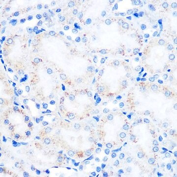
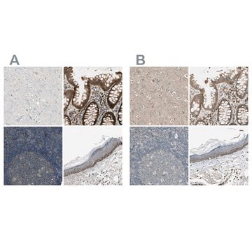
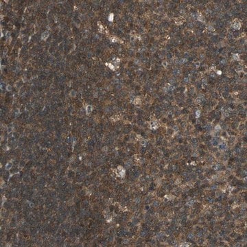
Anti-GLG1 antibody produced in rabbit, a Prestige Antibody, is developed and validated by the Human Protein Atlas (HPA) project . Each antibody is tested by immunohistochemistry against hundreds of normal and disease tissues. These images can be viewed on the Human Protein Atlas (HPA) site by clicking on the Image Gallery link. The antibodies are also tested using immunofluorescence and western blotting. To view these protocols and other useful information about Prestige Antibodies and the HPA, visit sigma.com/prestige.

Biochem/physiol Actions

GLG1 (golgi glycoprotein 1) binds to FGF (fibroblast growth factor), as well as to E-selectin and TGFβ (transforming growth factor β). It regulates the function of TGFβ, by modulating its maturation and secretion. This protein interacts with FGF18 at the cell surface, and positively regulates the FGF18 signaling cascade. GLG1 might act as a chaperone protein by regulating the processing and targeting of basic fibroblast growth factor (bFGF), in the cell. This protein is also associated with malignant astrocytomas in humans, and might have a role in the malignancy of these tumors. It is suggested that GLG1 aids in binding of E-selectin to myeloid cells. It might also play a role in the initiation of leukocyte endothelial contact formation, as it is expressed in microvilli of leukocytes.

Features and Benefits

Prestige Antibodies® are highly characterized and extensively validated antibodies with the added benefit of all available characterization data for each target being accessible via the Human Protein Atlas portal linked just below the product name at the top of this page. The uniqueness and low cross-reactivity of the Prestige Antibodies® to other proteins are due to a thorough selection of antigen regions, affinity purification, and stringent selection. Prestige antigen controls are available for every corresponding Prestige Antibody and can be found in the linkage section.

Every Prestige Antibody is tested in the following ways:
  • IHC tissue array of 44 normal human tissues and 20 of the most common cancer type tissues.
  • Protein array of 364 human recombinant protein fragments.

Linkage

Corresponding Antigen APREST71695

Physical form

Solution in phosphate-buffered saline, pH 7.2, containing 40% glycerol and 0.02% sodium azide

Legal Information

Prestige Antibodies is a registered trademark of Merck KGaA, Darmstadt, Germany

Disclaimer

Unless otherwise stated in our catalog or other company documentation accompanying the product(s), our products are intended for research use only and are not to be used for any other purpose, which includes but is not limited to, unauthorized commercial uses, in vitro diagnostic uses, ex vivo or in vivo therapeutic uses or any type of consumption or application to humans or animals.

Not finding the right product?  

Try our Product Selector Tool.

Storage Class Code

10 - Combustible liquids

WGK

WGK 1

Flash Point(F)

Not applicable

Flash Point(C)

Not applicable

Personal Protective Equipment

dust mask type N95 (US), Eyeshields, Gloves

Certificates of Analysis (COA)

Search for Certificates of Analysis (COA) by entering the products Lot/Batch Number. Lot and Batch Numbers can be found on a product’s label following the words ‘Lot’ or ‘Batch’.

Already Own This Product?

Find documentation for the products that you have recently purchased in the Document Library.

Visit the Document Library

Michaela C Baldauf et al.
Oncotarget, 9(2), 1587-1601 (2018-02-09)
Ewing sarcoma is an undifferentiated small-round-cell sarcoma. Although molecular detection of pathognomonic EWSR1-ETS fusions such as EWSR1-FLI1 enables definitive diagnosis, substantial confusion can arise if molecular diagnostics are unavailable. Diagnosis based on the conventional immunohistochemical marker CD99 is unreliable due
Fumio Yamaguchi et al.
International journal of oncology, 22(5), 1045-1049 (2003-04-10)
Malignant astrocytomas are highly invasive, vascular neoplasms that comprise the majority of nervous system tumors in humans. A strong association has previously been made between malignancy in human astrocytic tumors and increased expression of certain fibroblast growth factor (FGF) family
M Steegmaier et al.
Journal of cell science, 110 ( Pt 6), 687-694 (1997-03-01)
Neutrophils and subsets of lymphocytes bind to E-selectin, a cytokine inducible adhesion molecule on endothelial cells. The E-selectin-ligand-1 (ESL-1) is a high affinity glycoprotein ligand which participates in the binding of mouse myeloid cells to E-selectin. The sequence of mouse
M K Wild et al.
The Journal of biological chemistry, 276(34), 31602-31612 (2001-06-19)
E-selectin is an endothelial adhesion molecule, which mediates the tethering and rolling of leukocytes on vascular endothelium. It recognizes the glycoprotein E-selectin ligand-1 (ESL-1) as a major binding partner on mouse myeloid cells. Using surface plasmon resonance, we measured the
Jongcheol Ahn et al.
Journal of cell science, 118(Pt 8), 1725-1731 (2005-03-31)
The initial step in trafficking of leukocytes through the vascular endothelium is mediated by an adhesive interaction between molecules of the selectin family and their cognate receptors. Previously, a putative murine E-selectin ligand-1 (ESL-1) was identified and found to be

Our team of scientists has experience in all areas of research including Life Science, Material Science, Chemical Synthesis, Chromatography, Analytical and many others.

Contact Technical Service