Monoclonal Anti-Uvomorulin/E-Cadherin (rat IgG1 isotype) is derived from the DECMA-1 hybridoma, produced by the fusion of rat myeloma cells and splenocytes from an immunized Lou rat. Uvomorulin protein was initially identified in embryonal carcinoma and is identical to E-Cadherin, liver-cell adhesion molecules (L-CAM), Cell CAM 80/120, and Activity-regulated cytoskeleton-associated protein 1 (ARC-1), each of which have been characterized in different systems. Uvomorulin/E-Cadherin has been characterized as a 120 kDa cell surface glycoprotein from which an 84 kDa fragment can be released by trypsin digestion in the presence of Ca2+.
Specificity
Monoclonal Anti-Uvomorulin/E-Cadherin was selected against the mouse cell adhesion molecule uvomorulin/E-Cadherin. The antibody localizes the cell surface glycoprotein uvomorulin/E-cadherin that has been found to be identical to L-CAM, Cell CAM 80/120, and ARC-1. It blocks both the aggregation of mouse embryonal carcinoma cells and the compaction of pre-implantation embryos. The antibody disrupts confluent monolayers of Madin-Darby canine kidney (MDCK) epithelial cells. In indirect immunofluorescent staining of MDCK cells grown in culture, the antibody shows strong staining on the membrane of adjacent cells, after treatment with 0.5% Triton-X 100.
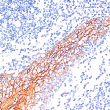
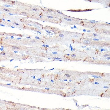
The antibody localizes the cell surface glycoprotein uvomorulin/E-cadherin that has been found to be identical to L-CAM, Cell CAM 80/120, and ARC-1. The antibody may be used for studies of embryonal development, cell-cell interactions of cultured cells, and localization of uvomorulin/E-cadherin in immunoblotting or immunohistochemical assays.
Immunogen
mouse embryonal carcinoma cell line PCC4 Aza R1.
Application
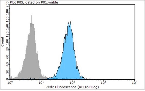
Monoclonal Anti-Uvomorulin/E-Cadherin has been used in immunofluorescence, immunoblotting, immunoprecipitation, immunohistochemistry, macromolecule permeability assay and agglomeration of two embryoid body assay.
Disclaimer
Unless otherwise stated in our catalog or other company documentation accompanying the product(s), our products are intended for research use only and are not to be used for any other purpose, which includes but is not limited to, unauthorized commercial uses, in vitro diagnostic uses, ex vivo or in vivo therapeutic uses or any type of consumption or application to humans or animals.
Epithelial morphogenesis involves a dramatic reorganisation of the microtubule cytoskeleton. How this complex process is controlled at the molecular level is still largely unknown. Here, we report that the centrosomal microtubule (MT)-binding protein CAP350 localises at adherens junctions in epithelial
The Journal of cell biology, 210(7), 1185-1197 (2015-09-30)
The first cell differentiation in mammalian embryos segregates polarized trophectoderm cells from an apolar inner cell mass (ICM). This lineage decision is specified in compacted morulae by cell polarization and adhesion acting on the Yes-associated protein in the Hippo signaling
Reduced Pax2 gene dosage increases apoptosis and slows the progression of renal cystic disease
Intratumoral heterogeneity and treatment resistance drive breast cancer (BC) metastasis and recurrence. The RUNX2 transcription factor is upregulated in early stage luminal BC. However, the precise mechanism by which RUNX2 regulates an oncogenic phenotype in luminal BCs remains an enigma.
Our team of scientists has experience in all areas of research including Life Science, Material Science, Chemical Synthesis, Chromatography, Analytical and many others.