


This antibody targets an epitope located in amino acid residues 4-10 at the N-terminus of the Opsin (rhodopsin) protein. As the retinal group is covalently linked to the opsin protein component on its lysine residues and the epitope region (AA 4-10) does not contain any lysine, it is unlikely that a retinal group is involved in the reactivity of this antibody. Therefore, the reactivity of this antibody would only involve the opsin component of the observed rhodopsin protein (UniProt#: P08100) upon labeling.
SAB4200762
Anti-Opsin antibody, Mouse monoclonal
clone RET-P1, purified from hybridoma cell culture
Synonym(s):
Anti-Opsin 2, Anti-Rhodopsin
Sign Into View Organizational & Contract Pricing
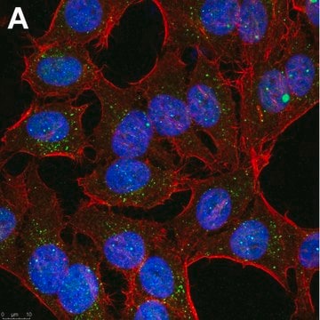
All Photos(2)
About This Item
UNSPSC Code:
12352203
NACRES:
NA.41
Pricing and availability is not currently available.
Recommended Products
biological source
mouse
Quality Level
antibody form
purified from hybridoma cell culture
antibody product type
primary antibodies
clone
RET-P1, monoclonal
form
buffered aqueous solution
mol wt
~39 kDa
species reactivity
avian, bovine, fish, mouse, rat, human, amphibian, turtle, rabbit
concentration
~1 mg/mL
technique(s)
ELISA: suitable
immunoblotting: 2-4 μg/mL using human retinoblastoma Y79 cell line extract.
immunofluorescence: suitable
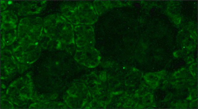
immunohistochemistry: 5-10 μg/mL using rat eye frozen sections.
radioimmunoassay: suitable
isotype
IgG1
UniProt accession no.
shipped in
dry ice
storage temp.
−20°C
target post-translational modification
unmodified
Gene Information
rat ... Rho(24717)
Related Categories
General description
Anti-Opsin antibody, Mouse monoclonal (rhodopsin) (mouse IgG1 isotype) is derived from the RET-P1 hybridoma produced by the fusion of mouse myeloma cells and splenocytes from BALB/C mice immunized with rat retinal membranes. Opsin is also called as rhodopsin protein, members of the G-protein coupled receptor (GPCR) superfamily. Opsin comprises >95% of the rod outer segments (ROS) intrinsic membrane and consists of a protein moiety - an opsin - and a non-protein moiety - the chromophore retinal.
Application
Anti-Opsin antibody has been used in
- immunoblotting
- immunofluorescence
- immunohistochemistry
- enzyme linked immunosorbent assay (ELISA)
- radioimmuno assay (RIA)
Biochem/physiol Actions
Opsin is an essential molecules for mediating the ability of animals to detect and use light for diverse biological functions. Mutations in the opsin (rhodopsin) gene are linked to retinitis pigmentosa (RP), a disease characterized by retinal degeneration resulting in reduced peripheral vision and night blindness.
Other Notes
rat retinal membranes
Physical form
Solution in 0.01 M phosphate buffered saline, pH 7.4, containing 15 mM sodium azide
Not finding the right product?
Try our Product Selector Tool.
Storage Class Code
10 - Combustible liquids
WGK
WGK 3
Flash Point(F)
Not applicable
Flash Point(C)
Not applicable
Choose from one of the most recent versions:
Certificates of Analysis (COA)
Lot/Batch Number
Don't see the Right Version?
If you require a particular version, you can look up a specific certificate by the Lot or Batch number.
Already Own This Product?
Find documentation for the products that you have recently purchased in the Document Library.
Retinitis pigmentosa
Hamel C.
Orphanet Journal of Rare Diseases, 1(1), 40-40 (2006)
The opsins
Terakita A.
Genome Biology, 6(3), 213-213 (2005)
-
I am confused by the wording in the description of the product on your website: "Anti-Opsin antibody, Mouse monoclonal (rhodopsin) (mouse IgG1 isotype) is derived from the RET-P1 hybridoma produced by the fusion of mouse myeloma cells and splenocytes from BALB/C mice immunized with rat retinal membranes. Opsin is also called as rhodopsin protein, members of the G-protein coupled receptor (GPCR) superfamily. Opsin comprises >95% of the rod outer segments (ROS) intrinsic membrane and consists of a protein moiety - an opsin - and a non-protein moiety - the chromophore retinal." Does this imply that this anti-opsin will recognize Rhodopsin as well, i.e. when we observe labeling for opsin using this antibody we could be observing Rhodopsin?
1 answer-
Helpful?
-
Active Filters
Our team of scientists has experience in all areas of research including Life Science, Material Science, Chemical Synthesis, Chromatography, Analytical and many others.
Contact Technical Service