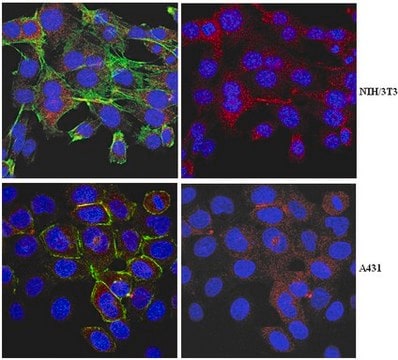
indirect immunofluorescence: 1:75 using cultured rat NRK cells microarray: suitable western blot: 1:1,000 using a whole extract of cultured dog MDCK cells
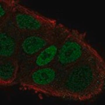
Myosin VI (MYO6) is localized to the Golgi complex and is expressed in the hair cells of the ear. It is a two-headed myosin with a short coiled-coil segment in its tail. The motor domain of MYO6 has two insertions. The gene encoding this protein is localized on chromosome 6q13.
Myosin VI is a relatively abundant widespread unconventional myosin composed of an N-terminal motor domain, a light chain binding neck region, a coiled-coiled region, and a highly conserved C-terminal domain. At the ′converter′ region, between the catalytic head and the neck region of Myosin VI, there is a characteristic linker about 50 amino acids long. Native Myosin VI is apparently a two-headed dimer of the heavy chains with each heavy chain bound to calmodulin light chain.
Immunogen
synthetic peptide corresponding to an epitope within the C-terminal of human Myosin VI, with N-terminal cysteine added, conjugated to KLH.
Myosin VI participates in the generation of cell shape change, cell motility, membrane remodeling, and possibly in organelle and particle transport or tethering. It is also involved in membrane trafficking pathways in cultured mammalian cells where it is associated with the membrane ruffles and the trans-Golgi network. The unusual direction of Myosin VI movement may suggest that it brings materials or membranes into the cell. Its activity in tissue cultured cells is thought to be regulated by phosphorylation. A mutation in Myosin VI was described recently in human autosomal dominant non syndromic hearing loss.
Physical form
Solution in 0.01 M phosphate buffered saline, pH 7.4, containing 1% BSA and 15 mM sodium azide.
Disclaimer
Unless otherwise stated in our catalog or other company documentation accompanying the product(s), our products are intended for research use only and are not to be used for any other purpose, which includes but is not limited to, unauthorized commercial uses, in vitro diagnostic uses, ex vivo or in vivo therapeutic uses or any type of consumption or application to humans or animals.
Individuals with Kabuki syndrome type 1 (KS1) often have hearing loss recognized in middle childhood. Current clinical dogma suggests that this phenotype is caused by frequent infections due to the immune deficiency in KS1 and/or secondary to structural abnormalities of
The Annals of otology, rhinology, and laryngology, 124 Suppl 1, 148S-157S (2015-05-23)
To elucidate the involvement of MYO6 mutations, known to be responsible for DFNA22/DFNB37, in Japanese hearing loss patients through the use of genetic analysis. Genomic variations responsible for hearing loss were identified by massively parallel DNA sequencing (MPS) of 63
Myosin VI is required for E-cadherin-mediated border cell migration
Aging leads to degeneration of the peripheral and central auditory systems, hearing loss, and difficulty understanding sounds in noise. Aging is also associated with changes in susceptibility to or recovery from damaging noise exposures, although the effects of the interaction
Our team of scientists has experience in all areas of research including Life Science, Material Science, Chemical Synthesis, Chromatography, Analytical and many others.