Skip to Content
Merck
All Photos(2)

Key Documents

05-537

Sigma-Aldrich

Anti-FAK Antibody, clone 4.47

clone 4.47, Upstate®, from mouse

Synonym(s):

Anti-FADK, Anti-FADK 1, Anti-FAK, Anti-FAK1, Anti-FRNK, Anti-PPP1R71, Anti-p125FAK, Anti-pp125FAK

Sign Into View Organizational & Contract Pricing


About This Item

UNSPSC Code:
12352203
eCl@ss:
32160702
NACRES:
NA.41

biological source

mouse

Quality Level

antibody form

purified immunoglobulin

antibody product type

primary antibodies

clone

4.47, monoclonal

species reactivity

mouse, rat, human

manufacturer/tradename

Upstate®

technique(s)

immunocytochemistry: suitable
immunohistochemistry: suitable
immunoprecipitation (IP): suitable
western blot: suitable

isotype

IgG1

NCBI accession no.

UniProt accession no.

shipped in

dry ice

target post-translational modification

unmodified

Gene Information

human ... PTK2(5747)

General description

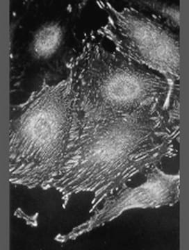
Focal adhesion kinase (FAK) is a cytoplasmic protein tyrosine kinase that has been implicated to play an important role in integrin-mediated signal transduction pathways. FAK becomes activated and tyrosine phosphorylated in integrin-mediated cell adhesion and is colocalized with integrins and other cytoskeletal proteins in focal contacts in a variety of adherent cells. Physical interactions of FAK with the integrin cytoplasmic domain as well as cytoskeletal proteins talin, paxillin, and/ or tensin have been proposed to play a key role in FAK activation by facilitating its oligomerization and transphosphorylation. FAK shows a rapid increase in tyrosine phosphorylation when stimulated by diverse signaling molecules, including those that regulate embryonic development, cell proliferation, migration and apoptosis, and efficient study methods are in high demand.

Specificity

Recognizes p125FAK; does not cross react with Pyk-2.

Immunogen

GST fusion protein corresponding to residues 1-423 of human FAK (focal adhesion kinase); specific for p125FAK

Application

Anti-FAK Antibody, clone 4.47 is an antibody against FAK for use in IC, IH, IP & WB.
Research Category
Signaling
Research Sub Category
Cytoskeletal Signaling

Quality

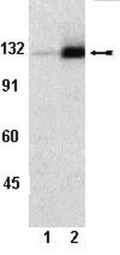
Routinely evaluated by immunoblot on 3T3/A31 RIPA cell lysate, PC-12, Hep-G2, SW 620 or Jurkat RIPA cell lysates

Target description

125 kDa

Linkage

Replaces: 04-591

Physical form

Format: Purified
Protein G Purified
Protein G Purified immunoglobulin in Immunoaffinity Purified immunoglobulin in 0.2M Tris-glycine, pH 7.4, 0.15M NaCl, 0.1mM EDTA, 0.05% sodium azide before the addition of glycerol to 30%

Storage and Stability

Maintain for 2 years at -20°C from date of shipment. Aliquot to avoid repeated freezing and thawing. For maximum recovery of product, centrifuge the original vial after thawing and prior to removing the cap.

Analysis Note

Control
Positive Antigen Control: Catalog #12-305, 3T3/A31 lysate. Add 2.5 μL of 2-mercapto-ethanol/100 μL of lysate and boil for 5 minutes to reduce the preparation. Load 20 μg of reduced lysate per lane for minigels.

Other Notes

Concentration: Please refer to the Certificate of Analysis for the lot-specific concentration.

Legal Information

UPSTATE is a registered trademark of Merck KGaA, Darmstadt, Germany

Disclaimer

Unless otherwise stated in our catalog or other company documentation accompanying the product(s), our products are intended for research use only and are not to be used for any other purpose, which includes but is not limited to, unauthorized commercial uses, in vitro diagnostic uses, ex vivo or in vivo therapeutic uses or any type of consumption or application to humans or animals.

Not finding the right product?  

Try our Product Selector Tool.

Storage Class Code

10 - Combustible liquids

WGK

WGK 1


Certificates of Analysis (COA)

Search for Certificates of Analysis (COA) by entering the products Lot/Batch Number. Lot and Batch Numbers can be found on a product’s label following the words ‘Lot’ or ‘Batch’.

Already Own This Product?

Find documentation for the products that you have recently purchased in the Document Library.

Visit the Document Library

B van de Water et al.
The Journal of biological chemistry, 274(19), 13328-13337 (1999-05-01)
The relationship between focal adhesion protein (FAK) activity and loss of cell-matrix contact during apoptosis is not entirely clear nor has the role of FAK in chemically induced apoptosis been studied. We investigated the status of FAK phosphorylation and cleavage
Y Shen et al.
Molecular biology of the cell, 10(8), 2507-2518 (1999-08-06)
The focal adhesion kinase (FAK) is discretely localized to focal adhesions via its C-terminal focal adhesion-targeting (FAT) sequence. FAK is regulated by integrin-dependent cell adhesion and can regulate tyrosine phosphorylation of downstream substrates, like paxillin. By the use of a
Sanjeev K Baniwal et al.
PloS one, 8(6), e62361-e62361 (2013-06-12)
Prolactin-induced Protein (PIP), an aspartyl protease unessential for normal mammalian cell function, is required for the proliferation and invasion of some breast cancer (BCa) cell types. Because PIP expression is particularly high in the Luminal A BCa subtype, we investigated
FAK inhibition decreases cell invasion, migration and metastasis in MYCN amplified neuroblastoma.
Megison, ML; Stewart, JE; Nabers, HC; Gillory, LA; Beierle, EA
Clinical & Experimental Metastasis null
FAK/PYK2 promotes the Wnt/?-catenin pathway and intestinal tumorigenesis by phosphorylating GSK3?.
Gao, C; Chen, G; Kuan, SF; Zhang, DH; Schlaepfer, DD; Hu, J
eLife null

Our team of scientists has experience in all areas of research including Life Science, Material Science, Chemical Synthesis, Chromatography, Analytical and many others.

Contact Technical Service