Saltar al contenido
Merck

M3807

Sigma-Aldrich

Anti-MAP Kinase, Non-Phosphorylated ERK antibody, Mouse monoclonal

clone ERK-NP2, purified from hybridoma cell culture

Sinónimos:

Anti-ERK, Anti-ERK-2, Anti-ERK2, Anti-ERT1, Anti-MAPK2, Anti-NS13, Anti-P42MAPK, Anti-PRKM1, Anti-PRKM2, Anti-p38, Anti-p40, Anti-p41, Anti-p41mapk, Anti-p42-MAPK

Iniciar sesiónpara Ver la Fijación de precios por contrato y de la organización


About This Item

MDL number:
UNSPSC Code:
12352203
NACRES:
NA.44

biological source

mouse

Quality Level

conjugate

unconjugated

antibody form

purified from hybridoma cell culture

antibody product type

primary antibodies

clone

ERK-NP2, monoclonal

form

buffered aqueous solution

mol wt

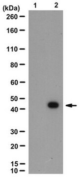
antigen, ERK-1 44 kDa
antigen, ERK-2 42 kDa

species reactivity

human, rat

concentration

~2 mg/mL

technique(s)

immunocytochemistry: suitable
indirect ELISA: suitable
microarray: suitable
western blot: 5-25 μg/mL using rat brain extract

isotype

IgG1

UniProt accession no.

shipped in

dry ice

storage temp.

−20°C

target post-translational modification

unmodified

Gene Information

General description

Mitogen-activated protein kinase (MAPK) superfamily of enzymes is involved in widespread signalling pathways. Members of this family include the ERK1/2 (extracellular signal-regulated protein kinase, also termed p42/p44 MAPK), JNK and p38 MAPK subfamilies. These are the terminal enzymes in a signalling cascade where each kinase phosphorylates and activates the next member in the sequence. Phosphorylation of both tyrosine and threonine is essential for the full activation of all MAPKs. Several kinases participate in activation of the ERK cascade. This cascade is initiated by the small G protein Ras, which upon stimulation causes activation Raf1 kinase. Raf1 continues the transmission by activating MEK. Activated MEK appears to be the only kinase capable of specifically phosphorylating and activating ERK. ERK appears to be an important regulatory molecule, which by can phosphorylate regulatory targets in the cytosol (phospholipase A2, PLA2), translocated into and phosphorylate substrates in the nucleus (ELK1). The activation of ERK cascade mediates and regulates the signal transduction pathways in response to stress, mitogenic signals and is important in development and differentiation, learning, memory and survival.

Specificity

The antibody reacts specifically with the non-phosphorylated, non-activated form of MAP kinase (ERK-1 and ERK-2). Weak cross-reaction is observed with monophosphorylated (threonine or tyrosine), but not with diphosphorylated peptides of MAPK. The antibody does not recognize JNK or p38 MAPK. The epitope recognized by the antibody contains non-phosphorylated threonine 183 and tyrosine 185 which resides within the ERK-activation loop (e.g., amino acids 178-188 in ERK-2).

Immunogen

synthetic peptide HTGFLTEYVAT, corresponding to the non-phosphorylated form of ERK-activation loop.

Application

A working concentration of 5-25 μg/mL may be used for detection by immunoblotting in rat brain extract. The antibody has also been used at working dilution of 1:50 for MAPK quantification by flow cytometry in monocyte-derived macrophages (MDM) derived from Holstein cattle.

Physical form

Solution in phosphate buffered saline, pH 7.4, containing 15 mM sodium azide.

Preparation Note

Prepared from a culture supernatant of bioreactor grown hybridoma

Disclaimer

Unless otherwise stated in our catalog or other company documentation accompanying the product(s), our products are intended for research use only and are not to be used for any other purpose, which includes but is not limited to, unauthorized commercial uses, in vitro diagnostic uses, ex vivo or in vivo therapeutic uses or any type of consumption or application to humans or animals.

¿No encuentra el producto adecuado?  

Pruebe nuestro Herramienta de selección de productos.

Storage Class

10 - Combustible liquids

wgk_germany

nwg

flash_point_f

Not applicable

flash_point_c

Not applicable


Certificados de análisis (COA)

Busque Certificados de análisis (COA) introduciendo el número de lote del producto. Los números de lote se encuentran en la etiqueta del producto después de las palabras «Lot» o «Batch»

¿Ya tiene este producto?

Encuentre la documentación para los productos que ha comprado recientemente en la Biblioteca de documentos.

Visite la Librería de documentos

Margarita Arango-Lievano et al.
Molecular and cellular biology, 36(6), 1019-1031 (2016-01-21)
Palmitoylation is involved in several neuropsychiatric and movement disorders for which a dysfunctional signaling of the dopamine D3 receptor (Drd3) is hypothesized. Computational modeling of Drd3's homologue, Drd2, has shed some light on the putative role of palmitoylation as a
Raffaella Molteni et al.
Neural plasticity, 2016, 2592319-2592319 (2016-02-18)
Although activity-dependent transcription represents a crucial mechanism for long-lasting experience-dependent changes in the hippocampus, limited data exist on its contribution to pathological conditions. We aim to investigate the influence of chronic stress on the activity-dependent transcription of brain-derived neurotrophic factor
Xin Wan et al.
Molecular medicine reports, 9(1), 229-234 (2013-10-23)
In this study, the role of the endothelin B receptor (ETBR) in oligodendroglioma cell proliferation and survival was investigated in vitro and in vivo. The overexpression and knockdown of ETBR was conducted in Hs683 human oligodendroglioma cells, and cell proliferation and activation (phosphorylation)
Michelle C Mendoza et al.
Trends in biochemical sciences, 36(6), 320-328 (2011-05-03)
The Ras-extracellular signal-regulated kinase (Ras-ERK) and phosphatidylinositol 3-kinase-mammalian target of rapamycin (PI3K-mTOR) signaling pathways are the chief mechanisms for controlling cell survival, differentiation, proliferation, metabolism, and motility in response to extracellular cues. Components of these pathways were among the first
Alexandra Fletcher-Jones et al.
eLife, 8 (2019-05-01)
Cannabinoid type one receptor (CB1R) is only stably surface expressed in axons, where it downregulates neurotransmitter release. How this tightly regulated axonal surface polarity is established and maintained is unclear. To address this question, we used time-resolved imaging to determine

Nuestro equipo de científicos tiene experiencia en todas las áreas de investigación: Ciencias de la vida, Ciencia de los materiales, Síntesis química, Cromatografía, Analítica y muchas otras.

Póngase en contacto con el Servicio técnico