Saltar al contenido
Merck
Todas las fotos(2)

Documentos clave

MABC292

Sigma-Aldrich

Anti-TAOK1 Antibody (long isoform), clone TAO1_1.2_VMD

clone 1.2, 1 mg/mL, from mouse

Sinónimos:

Serine/threonine-protein kinase TAO1, Kinase from chicken homolog B, hKFC-B, MARK Kinase, MARKK, Prostate-derived sterile 20-like kinase 2, PSK-2, PSK2, Prostate-derived STE20-like kinase 2, Thousand and one amino acid protein kinase 1, TAOK1, hTAOK1

Iniciar sesiónpara Ver la Fijación de precios por contrato y de la organización


About This Item

UNSPSC Code:
12352203
eCl@ss:
32160702
NACRES:
NA.41

biological source

mouse

Quality Level

antibody form

purified immunoglobulin

antibody product type

primary antibodies

clone

1.2, monoclonal

species reactivity

human

concentration

1 mg/mL

technique(s)

western blot: suitable

isotype

IgG2bκ

NCBI accession no.

UniProt accession no.

shipped in

wet ice

target post-translational modification

unmodified

Gene Information

human ... TAOK1(57551)

General description

TAOK1 , also known as Serine/threonine-protein kinase TAO1, Kinase from chicken homolog B, hKFC-B, MARK Kinase, MARKK, Prostate-derived sterile 20-like kinase 2, PSK-2/PSK2, Prostate-derived STE20-like kinase 2, or Thousand and one amino acid protein kinase 1, TAOK1/hTAOK1, and encoded by the gene TAOK1/KIAA1361/MAP3K16/MARKK, is an important serine/threonine protein kinase involved in a variety of processes including p38/MAPK14 stress responses, DNA damage responses and cytoskeleton stability. TAOK1 also acts as regulator of apoptosis and controls the morphological changes associated with apoptosis including cell contraction, membrane blebbing and apoptotic body formation. TAOK1 is expressed in many tissues but highly expressed in testis as well as brain, colon and muscle. TAOK1 is localized to the cytoplasm and a target for caspase-3 as well as SPRED1 a regulator of the MAP kinase cascade.

Immunogen

Epitope: C-terminus
Linear peptide corresponding to the C-terminus of human TAOK1.

Application

Research Category
Apoptosis & Cancer
Research Sub Category
Apoptosis - Additional
This Anti-TAOK1 Antibody (long isoform), clone TAO1_1.2_VMD is validated for use in Western Blotting for the detection of TAOK1 (long isoform).
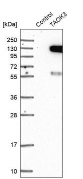
Western Blotting Analysis: A representative lot detected TAOK1 in HeLa cell lysate, but not in TAOK1 siRNA transfected HeLa cell lysate (Prof. Viji Draviam, University of Cambridge).

Quality

Evaluated by Western Blotting in HeLa cell lysate.

Western Blotting Analysis: 0.5 µg/mL of this antibody detected TAOK1 in 10 µg of HeLa cell lysate.

Target description

~116 kDa observed

Physical form

Format: Purified
Protein G Purified
Purified mouse monoclonal IgG2bκ in buffer containing 0.1 M Tris-Glycine (pH 7.4), 150 mM NaCl with 0.05% sodium azide.

Storage and Stability

Stable for 1 year at 2-8°C from date of receipt.

Disclaimer

Unless otherwise stated in our catalog or other company documentation accompanying the product(s), our products are intended for research use only and are not to be used for any other purpose, which includes but is not limited to, unauthorized commercial uses, in vitro diagnostic uses, ex vivo or in vivo therapeutic uses or any type of consumption or application to humans or animals.

¿No encuentra el producto adecuado?  

Pruebe nuestro Herramienta de selección de productos.

Storage Class

12 - Non Combustible Liquids

wgk_germany

WGK 1

flash_point_f

Not applicable

flash_point_c

Not applicable


Certificados de análisis (COA)

Busque Certificados de análisis (COA) introduciendo el número de lote del producto. Los números de lote se encuentran en la etiqueta del producto después de las palabras «Lot» o «Batch»

¿Ya tiene este producto?

Encuentre la documentación para los productos que ha comprado recientemente en la Biblioteca de documentos.

Visite la Librería de documentos

Nuestro equipo de científicos tiene experiencia en todas las áreas de investigación: Ciencias de la vida, Ciencia de los materiales, Síntesis química, Cromatografía, Analítica y muchas otras.

Póngase en contacto con el Servicio técnico