Skip to Content
Merck
All Photos(2)

Documents

MABN37

Sigma-Aldrich

Anti-Ataxin-1 Antibody, 11NQ, clone N76/8

clone N76/8, from mouse

Synonym(s):

ataxin 1, Spinocerebellar ataxia type 1 protein, spinocerebellar ataxia 1 (olivopontocerebellar ataxia 1, autosomal dominant, ataxin 1), ataxin-1

Sign Into View Organizational & Contract Pricing


About This Item

UNSPSC Code:
12352203
eCl@ss:
32160702
NACRES:
NA.41

biological source

mouse

Quality Level

antibody form

purified immunoglobulin

antibody product type

primary antibodies

clone

N76/8, monoclonal

species reactivity

rat

species reactivity (predicted by homology)

mouse (immunogen homology)

technique(s)

immunohistochemistry: suitable

isotype

IgG2bκ

NCBI accession no.

UniProt accession no.

shipped in

wet ice

target post-translational modification

unmodified

Gene Information

human ... ATXN1(6310)

General description

Ataxin-1 is a member of the ATXN1 protein family and contains a single AXH domain. It is a neurodegenerative disorder protein thought to have a role in the metabolism of RNA as it has been shown to localize to the RNA and transcription dependent inclusions within the nucleus. A mutation of Ataxin-1 is the cause of spinocerebellar ataxia type-1 (SCA1), a progressive, neurodegenerative disease that is autosomal dominant and primarily affects the Purjinke cells found in brain stem neuronal populations and the cerebellum. Expression of Ataxin-1 is almost ubiquitous, except in the brain where it is isolated to populations of neurons.

Immunogen

Linear peptide corresponding to mouse Ataxin-1.

Application

Anti-Ataxin-1 Antibody, 11NQ, clone N76/8 detects level of Ataxin-1 & has been published & validated for use in IH.
Research Category
Neuroscience
Research Sub Category
Neurodegenerative Diseases
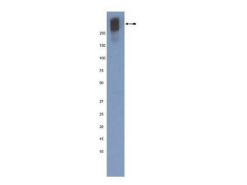
Western Blot Analysis: A previous lot was used by an independent laboratory in rat brain tissue lysate. (James Trimmer, UC Davis/NIH NeuroMab Facility)

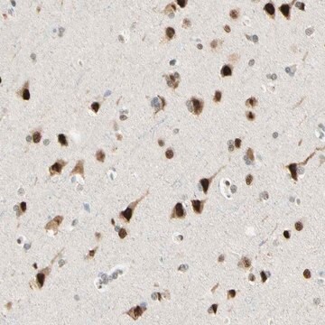
Immunohistochemistry Analysis: 1:500 dilution from a previous lot detected Ataxin-1 in rat cerebral cortex tissue.

Quality

Evaluated by Immunohistochemistry in rat hippocampus tissue.

Immunohistochemistry Analysis: 1:500 dilution of this antibody detected Ataxin-1 in rat hippocampus tissue.

Target description

~ 85 kDa observed

Physical form

Format: Purified
Protein G
Purified mouse monoclonal IgG2bκ in buffer containing 0.1 M Tris-Glycine (pH 7.4), 150 mM NaCl with 0.05% sodium azide.

Storage and Stability

Stable for 1 year at 2-8°C from date of receipt.

Analysis Note

Control
Rat hippocampus tissue

Other Notes

Concentration: Please refer to the Certificate of Analysis for the lot-specific concentration.

Disclaimer

Unless otherwise stated in our catalog or other company documentation accompanying the product(s), our products are intended for research use only and are not to be used for any other purpose, which includes but is not limited to, unauthorized commercial uses, in vitro diagnostic uses, ex vivo or in vivo therapeutic uses or any type of consumption or application to humans or animals.

Not finding the right product?  

Try our Product Selector Tool.

Storage Class Code

12 - Non Combustible Liquids

WGK

WGK 1

Flash Point(F)

Not applicable

Flash Point(C)

Not applicable


Certificates of Analysis (COA)

Search for Certificates of Analysis (COA) by entering the products Lot/Batch Number. Lot and Batch Numbers can be found on a product’s label following the words ‘Lot’ or ‘Batch’.

Already Own This Product?

Find documentation for the products that you have recently purchased in the Document Library.

Visit the Document Library

Juliana Bosso Taniguchi et al.
Human molecular genetics, 25(20), 4432-4447 (2017-02-09)
DNA damage and repair is a critical domain of many neurodegenerative diseases. In this study, we focused on RpA1, a candidate key molecule in polyQ disease pathologies, and tested the therapeutic effect of adeno-associated virus (AAV) vector expressing RpA1 on
Sam S Barclay et al.
Human molecular genetics, 23(5), 1345-1364 (2013-11-02)
DNA damage repair is implicated in neurodegenerative diseases; however, the relative contributions of various DNA repair systems to the pathology of these diseases have not been investigated systematically. In this study, we performed a systematic in vivo screen of all
Kyota Fujita et al.
Nature communications, 8(1), 1864-1864 (2017-12-02)
YAP and its neuronal isoform YAPdeltaC are implicated in various cellular functions. We found that expression of YAPdeltaC during development, but not adulthood, rescued neurodegeneration phenotypes of mutant ataxin-1 knock-in (Atxn1-KI) mice. YAP/YAPdeltaC interacted with RORα via the second WW

Our team of scientists has experience in all areas of research including Life Science, Material Science, Chemical Synthesis, Chromatography, Analytical and many others.

Contact Technical Service