SAB5500002
Anti-SMA antibody, Rabbit monoclonal
clone SP171, recombinant, expressed in proprietary host, affinity isolated antibody
Sign Into View Organizational & Contract Pricing
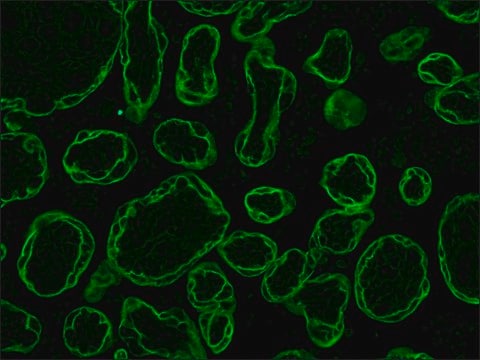
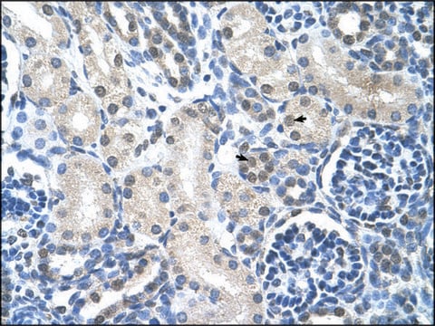
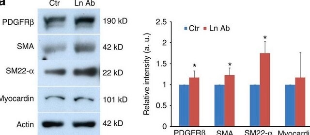
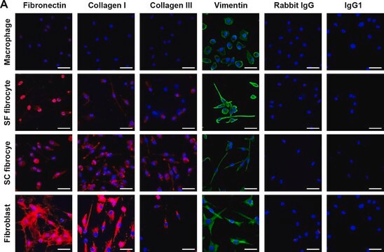
All Photos(2)
About This Item
UNSPSC Code:
12352203
NACRES:
NA.41
conjugate:
unconjugated
application:
IHC
clone:
SP171, monoclonal
species reactivity:
human (tested)
citations:
28
technique(s):
immunoblotting: 1:50
immunohistochemistry: 1:200
immunohistochemistry: 1:200
Recommended Products
biological source
rabbit
Quality Level
recombinant
expressed in proprietary host
conjugate
unconjugated
antibody form
affinity isolated antibody
antibody product type
primary antibodies
clone
SP171, monoclonal
species reactivity
human (tested)
species reactivity (predicted by homology)
rabbit, rat, bovine, chicken, mouse, pig
technique(s)
immunoblotting: 1:50
immunohistochemistry: 1:200
isotype
IgG
UniProt accession no.
shipped in
wet ice
storage temp.
2-8°C
target post-translational modification
unmodified
Gene Information
human ... ACTA2(59)
General description
Smooth muscle actin-α (SMA), also known as α2-smooth muscle actin (ACTA2), is a cytoskeleton protein in smooth muscle cells. It is encoded by the gene mapped to human chromosome 10q23.31. SMA is a vascular smooth muscle specific isoform,
Smooth muscle actin-alpha (SMA) is a cytoskeleton protein in smooth muscle cells and their derived tumors such as leiomyoma and leiomyosarcoma. It is also expressed in myoepithelial cells of the breast and salivary gland, but not in fibroblasts, striated muscle, and myocardium.
Immunogen
Synthetic peptide near the N-terminus of human SMA protein.
Application
Anti-SMA antibody, Rabbit monoclonal has been used in:
- western blotting
- immunohistochemistry
- immunofluorescence
Biochem/physiol Actions
Smooth muscle actin-α (SMA)/ α2-smooth muscle actin (ACTA2) interacts with β-myosin heavy chain and facilitates vascular smooth muscle cell contraction. The encoded protein regulates c-MET (tyrosine-protein kinase Met) and focal adhesion kinase (FAK) expression in human lung adenocarcinoma cells, which positively and selectively mediates tumor progression. Thus, SMA can be used as a potential prognostic biomarker and/or target for treating metastatic lung adenocarcinoma. Mutation in the gene is associated with the development of patent ductus arteriosus (PDA), bicuspid aortic valve (BAV), iris flocculi, livedo reticularis, cerebrovascular accident (CVA) and stenosis of the aortic vasa vasorum. In addition, variation in the gene expression leads to thoracic aortic aneurysms and dissections (TAAD).
Features and Benefits
Evaluate our antibodies with complete peace of mind. If the antibody does not perform in your application, we will issue a full credit or replacement antibody. Learn more.
Physical form
0.1 ml rabbit monoclonal antibody purified by protein A/G in PBS/1% BSA buffer pH 7.6 with less than 0.1% sodium azide.
Disclaimer
Unless otherwise stated in our catalog or other company documentation accompanying the product(s), our products are intended for research use only and are not to be used for any other purpose, which includes but is not limited to, unauthorized commercial uses, in vitro diagnostic uses, ex vivo or in vivo therapeutic uses or any type of consumption or application to humans or animals.
Not finding the right product?
Try our Product Selector Tool.
Storage Class Code
10 - Combustible liquids
WGK
WGK 2
Flash Point(F)
Not applicable
Flash Point(C)
Not applicable
Choose from one of the most recent versions:
Already Own This Product?
Find documentation for the products that you have recently purchased in the Document Library.
Customers Also Viewed
Ying-Chun Zhu et al.
International journal of molecular medicine, 40(4), 1165-1171 (2017-08-30)
Transforming growth factor-β (TGF-β) induces epithelial-mesenchymal transition (EMT) primarily via a Smad‑dependent mechanism. However, there are few studies available on TGF-β-induced EMT through the activation of non‑canonical pathways. In this study, the Cdc42-interacting protein-4 (CIP4)/partitioning-defective protein 6 (Par6) pathway was investigated in TGF-β1‑stimulated NRK-52E cells. Rat
The genetics and genomics of thoracic aortic disease.
Pomianowski P and John A E
Journal of Cardiothoracic Surgery, 2(3), 271-271 (2013)
Suppression of CIP4/Par6 attenuates TGF-β1-induced epithelial-mesenchymal transition in NRK-52E cells.
Zhu Y-C, et al.
International Journal of Molecular Medicine, 40(4), 1165-1171 (2017)
BBC3 in macrophages promoted pulmonary fibrosis development through inducing autophagy during silicosis.
Liu H, et al.
Cell Death & Disease, 8(3), e2657-e2657 (2017)
Macrophage-derived MCPIP1 mediates silica-induced pulmonary fibrosis via autophagy.
Liu H, et al.
Particle and Fibre Toxicology, 13(1), 55-55 (2016)
Our team of scientists has experience in all areas of research including Life Science, Material Science, Chemical Synthesis, Chromatography, Analytical and many others.
Contact Technical Service


