Skip to Content
Merck
All Photos(8)

Key Documents

C6219

Sigma-Aldrich

Anti-Connexin 43 Antibody

rabbit polyclonal

Synonym(s):

Connexin 43 Antibody, Connexin 43 Antibody - Anti-Connexin-43 antibody produced in rabbit, Cx43 Antibody

Sign Into View Organizational & Contract Pricing


About This Item

MDL number:
UNSPSC Code:
12352203
NACRES:
NA.41
conjugate:
unconjugated
application:
ARR
ICC
IF
IHC (f)
IHC (p)
WB
clone:
polyclonal
species reactivity:
mammals, human
citations:
512
technique(s):
immunocytochemistry: suitable
immunohistochemistry (formalin-fixed, paraffin-embedded sections): 1:2,000 using human or animal tissue sections
immunohistochemistry (frozen sections): 1:2,000 using human or animal tissue sections
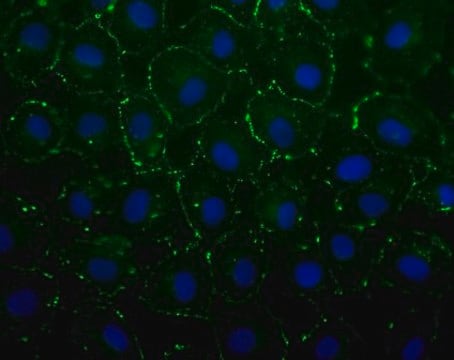
indirect immunofluorescence: 1:400 using cultured BHK cells
microarray: suitable
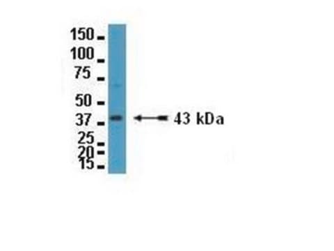
western blot: 1:8,000 using an animal tissue extract

Product Name

Anti-Connexin-43 antibody produced in rabbit, affinity isolated antibody, buffered aqueous solution

biological source

rabbit

Quality Level

conjugate

unconjugated

antibody form

affinity isolated antibody

antibody product type

primary antibodies

clone

polyclonal

form

buffered aqueous solution

species reactivity

mammals, human

packaging

antibody small pack of 25 μL

technique(s)

immunocytochemistry: suitable
immunohistochemistry (formalin-fixed, paraffin-embedded sections): 1:2,000 using human or animal tissue sections
immunohistochemistry (frozen sections): 1:2,000 using human or animal tissue sections
indirect immunofluorescence: 1:400 using cultured BHK cells
microarray: suitable
western blot: 1:8,000 using an animal tissue extract

UniProt accession no.

shipped in

dry ice

storage temp.

−20°C

target post-translational modification

unmodified

Gene Information

human ... GJA1(2697)
mouse ... Gja1(14609)
rat ... Gja1(24392)

General description

Anti-Connexin 43 is developed in rabbit using a synthetic peptide conjugated to KLH with glutaraldehyde as immunogen. The peptide corresponds to a C-terminal segment of the cytoplasmic domain of human and rat connexin 43, amino acid residues, with an N-terminal added lysine. Affinity isolated antigen specific antibody is obtained by immunospecific purification which removes essentially all rabbit serum proteins, including immunoglobulins, which do not specifically bind to connexin 43.

Gap junctions are specialized cell membrane domains consisting of aggregations of intercellular channels that directly connect the cytoplasm of adjacent cells. Gap junctions coordinate cellular and organ function in tissues and are involved in metabolic cooperation between cells, electrical coupling, synchronization of cellular physiological activities, growth control, and developmental regulation. The gap junction channels allow intercellular exchange of ions, nucleotides and small molecules between adjacent cells. Unlike other membrane channels, intercellular channels span two opposed plasma membranes and require the contribution of hemi-channels, called connexons, from both participating cells. These channels are permeable to molecules as large as 1 kDa, and they have been reported in most mammalian cell types. Two connexons interact in the extracellular space to form the complete intercellular channel. Each connexon is composed of six similar or identical proteins, which have been termed connexins. Connexins (Cx) are a multi-gene family of highly related proteins with molecular weights ranging from 26 to 70 kDa. At least a dozen distinct connexin genes have been identified in mammals, many expressed in a diverse tissue and cell specific pattern. Two distinct lineages have been identified in mammals, one termed class I or β group, in which Cx26, Cx30, Cx31, Cx31.1 and Cx32 fall, and the other termed class II or α group, represented by Cx33, Cx37, Cx40, Cx43 and Cx46.

Connexin-43 (Cx-43), a 43kDa protein, is a member of the connexin protein family that forms a gap junction. It is expressed by a variety of cell types, such as astrocytes, cardiac and smooth muscle, endothelium, ependyma, fibroblasts, keratinocytes, lens and corneal epithelium. In addition, the protein is also found in leptomeninges, leucocytes, Leydig cells, macrophages, myoepithelial cells of mammary gland, osteocytes, ovarian granulosa, pancreatic β-cells, preimplantation blastocyst, sertoli cells, thyroid follicular cells and trophoblast giant cells.
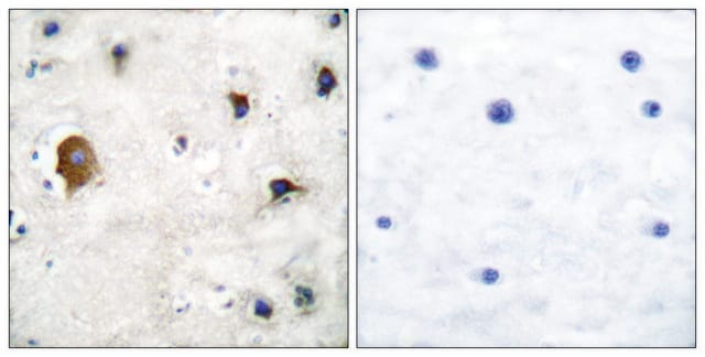
The 43 kDa connexin protein (connexin 43, Cx43) is a phosphoprotein that is expressed in most tissues, even though the pattern of expression may differ in various cell types (e.g., in the brain, it is found in astrocytes, ependyma and leptomeninges, but not in neurons, oligodendrocytes and pinealocytes; or in the liver, it is present in Ito cells, but not in hepatocytes). Gap junction protein levels change in response to disruption of tissue architecture. For instance, an increased expression of Cx43 was found in early stages of atherosclerosis.

Specificity

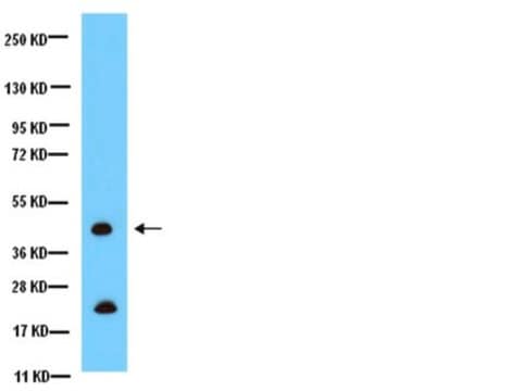
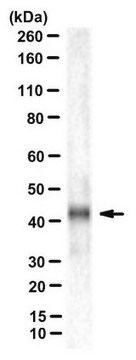
Anti-Connexin 43 reacts specifically with connexin 43. By immunoblotting, the antibody detects a single band or 2-3 bands at 43 kDa region. Staining of connexin 43 band(s) by immunoblotting is specifically inhibited with the connexin 43 peptide. Reactivity has been observed with human, bovine, rat, mouse, hamster and chicken connexin 43.

Immunogen

Synthetic peptide corresponding to the C-terminal segment of the cytoplasmic domain (amino acids with N-terminally added lysine) of human/rat connexin-43.

Application

Anti-Connexin-43 may be used in immunoblotting, immunocytochemistry and immunohistochemistry (frozen and formalin-fixed, paraffin-embedded tissues). Polyclonal antibodies reacting specifically with Cx43 may be applied in diverse cellular and molecular approaches to the study of gap junctions and their properties.
A minimum working dilution of 1:8,000 is determined by immunoblotting using a whole extract from mouse brain.
A minimum working dilution of 1:400 is determined by indirect immunofluorescent staining of acetone-fixed cultured baby hamster kidney (BHK).
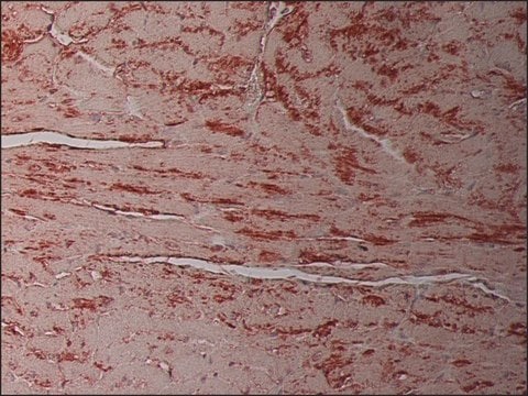
A minimum working dilution of 1:2,000 is determined by indirect immunofluorescent staining of rat heart. (Negative on rat liver sections)
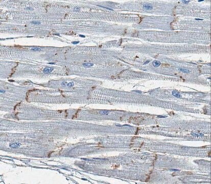
A minimum working dilution of 1:2,000 is determined by indirect immunoperoxidase staining of trypsin-digested,formalin-fixed, paraffin-embedded human or animal tissue.

Biochem/physiol Actions

Connexin-43 (Cx-43) might play a vital role in neuronal migration as it resides in radial glial fibers which are closely associated with migrating neurons. It is also involved in transmembrane NAD+ transport. Connexin-43, which is an element of gap junction, facilitates cell-cell communication. Cx-43 is known to induce [Ca2+]i burst phenomenon in uterine artery endothelial cells (UAECs). Elevated expression of Cx-43 has been observed during early stages of human coronary atherosclerosis. Reduction or alteration in the levels or types of connexin expressed in various cell types correlates with tumor progression and metastasis. However, glioma cells transfected with the oncogene neu (c-erb-B2) exhibit a major reduction in intercellular communication with no decrease in overall expression of Cx43. Different states of rat Cx43 phosphorylation are found during development and lactation. Connexin 43 mutations occur in children whose hearts failed to develop asymmetry and lethal heart malformations occur on knocking out this gene in mice. Polyclonal antibodies reacting specifically with Cx438-11 may be applied in diverse cellular and molecular approaches to the study of gap junctions and their properties, and to correlate their expression pattern in a variety of cell types and tissues with physiological functions or pathological conditions.

Physical form

Solution in 0.01 M phosphate buffered saline, pH 7.4, containing 1% bovine serum albumin and 15 mM sodium azide.

Storage and Stability

For continuous use, store at 2-8°C for up to one month. For extended storage freeze in working aliquots. Repeated freezing and thawing is not recommended. Storage in "frost-free" freezers is not recommended. If slight turbidity occurs upon prolonged storage, clarify the solution by centrifugation before use.

Disclaimer

Unless otherwise stated in our catalog or other company documentation accompanying the product(s), our products are intended for research use only and are not to be used for any other purpose, which includes but is not limited to, unauthorized commercial uses, in vitro diagnostic uses, ex vivo or in vivo therapeutic uses or any type of consumption or application to humans or animals.

Not finding the right product?  

Try our Product Selector Tool.

Storage Class Code

12 - Non Combustible Liquids

WGK

nwg

Flash Point(F)

Not applicable

Flash Point(C)

Not applicable


Choose from one of the most recent versions:

Certificates of Analysis (COA)

Lot/Batch Number

Don't see the Right Version?

If you require a particular version, you can look up a specific certificate by the Lot or Batch number.

Already Own This Product?

Find documentation for the products that you have recently purchased in the Document Library.

Visit the Document Library

Connexin 43 hemi channels mediate Ca2+-regulated transmembrane NAD+ fluxes in intact cells.
Bruzzone S, et al.
Faseb Journal, 15(1), 10-12 (2001)
Zonula occludens-1 alters connexin43 gap junction size and organization by influencing channel accretion.
Hunter A W, et al.
Molecular Biology of the Cell, 16(12), 5686-5698 (2005)
Naphichaya Phacharapiyangkul et al.
International journal of medical sciences, 16(8), 1107-1115 (2019-09-17)
Astragalus membranaceus has been shown to possess anti-inflammation and antitumor properties. Several studies have indicated that extracts of Astragalus membranaceus (PG2) have growth inhibitory effects on tumor. However, the effect of PG2 on enhancing the chemotherapy, modulating tumor immune escape
Gap junctions in the brain: where, what type, how many and why?
Dermietzel R and Spray D C.
Trends in Neurosciences, 16(5), 186-192 (1993)
Pregnancy enhances sustained Ca2+ bursts and endothelial nitric oxide synthase activation in ovine uterine artery endothelial cells through increased connexin 43 function.
Yi F X, et al.
Biology of Reproduction, 82(1), 66-75 (2010)

Articles

Cancer research has revealed that the classical model of carcinogenesis, a three step process consisting of initiation, promotion, and progression, is not complete.

Our team of scientists has experience in all areas of research including Life Science, Material Science, Chemical Synthesis, Chromatography, Analytical and many others.

Contact Technical Service