Skip to Content
Merck
All Photos(4)

Key Documents

C3678

Sigma-Aldrich

Anti-Pan Cadherin antibody produced in rabbit

whole antiserum

Synonym(s):

Anti-ACOGS, Anti-ARVD14, Anti-CD325, Anti-CDHN, Anti-CDw325, Anti-NCAD

Sign Into View Organizational & Contract Pricing


About This Item

MDL number:
UNSPSC Code:
12352203
NACRES:
NA.41
conjugate:
unconjugated
application:
IF
IHC (p)
WB
clone:
polyclonal
species reactivity:
rabbit, cat, chicken, human
citations:
55
technique(s):
immunohistochemistry (formalin-fixed, paraffin-embedded sections): 1:1,000 using protease-digested animal heart sections
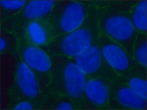
indirect immunofluorescence: 1:100 using cultured MDBK cells
western blot: 1:200

biological source

rabbit

Quality Level

conjugate

unconjugated

antibody form

whole antiserum

antibody product type

primary antibodies

clone

polyclonal

mol wt

antigen 135 kDa

contains

15 mM sodium azide

species reactivity

rabbit, cat, chicken, human

technique(s)

immunohistochemistry (formalin-fixed, paraffin-embedded sections): 1:1,000 using protease-digested animal heart sections
indirect immunofluorescence: 1:100 using cultured MDBK cells
western blot: 1:200

UniProt accession no.

shipped in

dry ice

storage temp.

−20°C

target post-translational modification

unmodified

Gene Information

chicken ... CDH2(414745)

General description

Cadherins are glycoprotein receptors that regulate Ca2+-dependant adhesion in cells. Cadherins are associated with adherens junctions and are mainly involved in morphogenesis of cells and tissues. P-cadherin is expressed in epithelial cells and its over-expression has been associated with breast cancer metastasis and invasion . Anti-Pan Cadherin antibody is specific for cadherins in a wide range of organisms including chickens, rabbits and rats.

Specificity

The epitope recognized by the antibody is shared by a wide variety of organisms.

Immunogen

Synthetic peptide corresponding to the C-terminal amino acids of chicken N-Cadherin with an extra N-terminal lysine residue (24 amino acids) coupled with glutaraldehyde to keyhole limpet hemocyanin (KLH).

Application

Anti-Pan Cadherin antibody is suitable for use in confocal immunofluorescence using rat ventricular myocytes and western blot (1:200). The antibody may also be used for indirect immunofluorescence (1:100 using cultured MDBK cells) and immunohistochemistry (in formalin-fixed, paraffin-embedded sections or at 1:1,000 dilutions using protease-digested animal heart sections). Additionally, the product can be used in indirect immunoblot (135 kDa band in chicken or rabbit heart extracts), immunoperoxidase and immunoelectron-microscopy assays.
May be used for the detection of members of the cadherin family or genetically engineered proteins containing the C-terminal cadherin tail, and for demonstration of adherens type cell-cell junctions regardless of their cadherin type.

Disclaimer

Unless otherwise stated in our catalog or other company documentation accompanying the product(s), our products are intended for research use only and are not to be used for any other purpose, which includes but is not limited to, unauthorized commercial uses, in vitro diagnostic uses, ex vivo or in vivo therapeutic uses or any type of consumption or application to humans or animals.

Not finding the right product?  

Try our Product Selector Tool.

Storage Class Code

12 - Non Combustible Liquids

WGK

nwg

Flash Point(F)

Not applicable

Flash Point(C)

Not applicable


Choose from one of the most recent versions:

Certificates of Analysis (COA)

Lot/Batch Number

Don't see the Right Version?

If you require a particular version, you can look up a specific certificate by the Lot or Batch number.

Already Own This Product?

Find documentation for the products that you have recently purchased in the Document Library.

Visit the Document Library

André Albergaria et al.
The International journal of developmental biology, 55(7-9), 811-822 (2011-12-14)
P-cadherin is a cell-cell adhesion molecule, whose expression is highly associated with undifferentiated cells in normal adult epithelial tissues, as well as with poorly differentiated carcinomas. Its expression has been already reported in human embryonic stem cells and it is
Kiyomi Yamada et al.
Circulation research, 97(4), 346-353 (2005-07-23)
To define mechanisms regulating expression of cell-cell junction proteins, we have developed an in vitro system in which neonatal rat ventricular myocytes were subjected to pulsatile stretch. Previously, we showed that expression of the gap junction protein, connexin (Cx) 43
Heyjin Lee et al.
International journal of molecular sciences, 18(12) (2017-12-21)
Cachexia and sarcopenia are the main causes of muscle atrophy. These result in a reduction in the muscle fiber area, myo-protein content, and muscle strength, with various molecular modulators being involved. Although several reports have proposed potential therapeutic agents, no
Sang-Jin Lee et al.
Cell death & disease, 7(10), e2431-e2431 (2016-10-21)
Skeletal myogenesis is coordinated by multiple signaling pathways that control cell adhesion/migration, survival and differentiation accompanied by muscle-specific gene expression. A cell surface protein Cdo is involved in cell contact-mediated promyogenic signals through activation of p38MAPK and AKT. Protein kinase
Laura Pelz et al.
The Journal of biological chemistry, 292(52), 21490-21503 (2017-11-11)
The Ig-like cell adhesion molecule (IgCAM) BT-IgSF (brain- and testis-specific Ig superfamily protein) plays a major role in male fertility in mice. However, the molecular mechanism by which BT-IgSF supports fertility is unclear. Here, we found that it is localized

Our team of scientists has experience in all areas of research including Life Science, Material Science, Chemical Synthesis, Chromatography, Analytical and many others.

Contact Technical Service