Skip to Content
Merck
All Photos(2)

Key Documents

MABT134

Sigma-Aldrich

Anti-VE-cadherin Antibody, clone BV6

clone BV6, from mouse

Synonym(s):

Cadherin-5, 7B4 Antigen

Sign Into View Organizational & Contract Pricing


About This Item

UNSPSC Code:
12352203
eCl@ss:
32160702
NACRES:
NA.41
clone:
BV6, monoclonal
application:
FACS
ICC
IHC
WB
species reactivity:
human
technique(s):
flow cytometry: suitable
immunocytochemistry: suitable
immunohistochemistry: suitable (paraffin)
western blot: suitable
citations:
13

biological source

mouse

Quality Level

antibody form

purified immunoglobulin

antibody product type

primary antibodies

clone

BV6, monoclonal

species reactivity

human

technique(s)

flow cytometry: suitable
immunocytochemistry: suitable
immunohistochemistry: suitable (paraffin)
western blot: suitable

isotype

IgG2aκ

NCBI accession no.

UniProt accession no.

shipped in

wet ice

target post-translational modification

unmodified

Gene Information

human ... CDH5(1003)

General description

Human Vascular Endothelial (VE)-cadherin is a calcium-dependent adhesion molecule strictly located at cell-to-cell junctions. VE-cadherin is present in all types of endothelium (veins, arteries, capillary and large vessels). Cadherins are calcium dependent cell adhesion proteins. They preferentially interact with themselves in a homophilic manner in connecting cells. Cadherins may thus contribute to the sorting of heterogeneous cell types. VE-cadherin may play an important role in endothelial cell biology through control of the cohesion and organization of the intercellular junctions. VE-cadherin also associates with alpha-catenin forming a link to the cytoskeleton.

Immunogen

HUVEC cells

Application

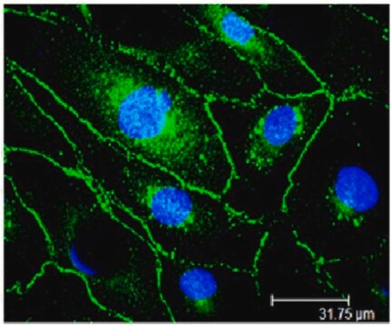
Immunocytochemistry Analysis: A 1:500 dilution from a representative lot detected VE-cadherin in the cell-cell junctions of HUVEC cells.

Immunohistochemistry Analysis: A 1:10-1:20 dilution of a representative lot was used by an independent laboratory in paraffin-embedded tissue sections. Buffered formalin fixation is recommended with a fixation period of no longer than 12 hours. High heat antigen retrieval in citrate buffer (Cat. No. 21545) is also suggested. Antibody can also be used to label acetone-fixed cryostat sections or cells using a immunoperoxidase staining protocol (eg. IHC Select Detection Kit, Cat. No. DAB150)

Flow Cytometry Analysis: A representative lot was used in the presence of Ca2+. Note: Use PBS + 2-5mM EDTA for cell detachment.

Western Blot Analysis: Non-reducing conditions may be needed, Ca2+ required in buffer (2-5 mM).
Research Category
Cell Structure
Research Sub Category
Adhesion (CAMs)
This Anti-VE-cadherin Antibody, clone BV6 is validated for use in WB, IC, FC, IH(P) for the detection of VE-cadherin.

Quality

Evaluated by Western Blot in HUVEC cell lysate.

Western Blot Anlaysis: A 1:2,000 dilution of this antibody detected VE-cadherin in HUVEC cell lysate.

Target description

~120 kDa observed.
The calculated molecular weight is 82 kDa but it will run between ~90-140 kDa because this protein is glycosolated.

Linkage

Replaces: MAB1989

Physical form

Format: Purified
Protein G Purified
Purified mouse monoclonal IgG2aκ in buffer containing 0.1 M Tris-Glycine (pH 7.4), 150 mM NaCl with 0.05% sodium azide.

Storage and Stability

Stable for 1 year at 2-8°C from date of receipt.

Analysis Note

Control
HUVEC cell lysate

Other Notes

Concentration: Please refer to the Certificate of Analysis for the lot-specific concentration.

Disclaimer

Unless otherwise stated in our catalog or other company documentation accompanying the product(s), our products are intended for research use only and are not to be used for any other purpose, which includes but is not limited to, unauthorized commercial uses, in vitro diagnostic uses, ex vivo or in vivo therapeutic uses or any type of consumption or application to humans or animals.

Not finding the right product?  

Try our Product Selector Tool.

Storage Class Code

12 - Non Combustible Liquids

WGK

WGK 1

Flash Point(F)

Not applicable

Flash Point(C)

Not applicable


Certificates of Analysis (COA)

Search for Certificates of Analysis (COA) by entering the products Lot/Batch Number. Lot and Batch Numbers can be found on a product’s label following the words ‘Lot’ or ‘Batch’.

Already Own This Product?

Find documentation for the products that you have recently purchased in the Document Library.

Visit the Document Library

Florian A Gegenfurtner et al.
Journal of cell science, 131(10) (2018-05-05)
Developmental processes, such as angiogenesis, are associated with a constant remodeling of the actin cytoskeleton in response to different mechanical stimuli. The mechanosensitive transcription factors MRTF-A (MKL1) and YAP (also known as YAP1) are important mediators of this challenging adaptation
Noritoshi Nagaya et al.
Circulation, 108(7), 889-895 (2003-07-02)
Circulating endothelial progenitor cells (EPCs) migrate to injured vascular endothelium and differentiate into mature endothelial cells. We investigated whether transplantation of vasodilator gene-transduced EPCs ameliorates monocrotaline (MCT)-induced pulmonary hypertension in rats. We obtained EPCs from cultured human umbilical cord blood
Yi Jin et al.
Nature cardiovascular research, 1(12), 1156-1173 (2023-11-08)
Vascular endothelial (VE)-cadherin in endothelial adherens junctions is an essential component of the vascular barrier, critical for tissue homeostasis and implicated in diseases such as cancer and retinopathies. Inhibitors of Src cytoplasmic tyrosine kinase have been applied to suppress VE-cadherin
Pan Liu et al.
Biotechnology and bioengineering, 118(1), 423-432 (2020-09-25)
Vascular leak is a key driver of organ injury in diseases, and strategies that reduce enhanced permeability and vascular inflammation are promising therapeutic targets. Activation of the angiopoietin-1 (ANG1)-Tie2 tyrosine kinase signaling pathway is an important regulator of vascular quiescence.
Maibritt Kretschmer et al.
Journal of cell science, 136(2) (2023-02-01)
Notch signaling is critical for many developmental and disease-related processes. It is widely accepted that Notch has a mechanotransduction module that regulates receptor cleavage. However, the role of biomechanical properties of the cellular environment in Notch signaling in general is

Our team of scientists has experience in all areas of research including Life Science, Material Science, Chemical Synthesis, Chromatography, Analytical and many others.

Contact Technical Service