Skip to Content
Merck
All Photos(1)

Key Documents

MAB1273

Sigma-Aldrich

Anti-Mitochondria Antibody, surface of intact mitochondria, clone 113-1

clone 113-1, Chemicon®, from mouse

Synonym(s):

Mitochondrial antibody

Sign Into View Organizational & Contract Pricing


About This Item

UNSPSC Code:
12352203
eCl@ss:
32160702
NACRES:
NA.41

biological source

mouse

Quality Level

antibody form

purified immunoglobulin

antibody product type

primary antibodies

clone

113-1, monoclonal

species reactivity

human

should not react with

mouse, rat

manufacturer/tradename

Chemicon®

technique(s)

immunofluorescence: suitable
immunohistochemistry (formalin-fixed, paraffin-embedded sections): suitable
immunoprecipitation (IP): suitable

input

sample type neural stem cell(s)

isotype

IgG1

shipped in

dry ice

target post-translational modification

unmodified

General description

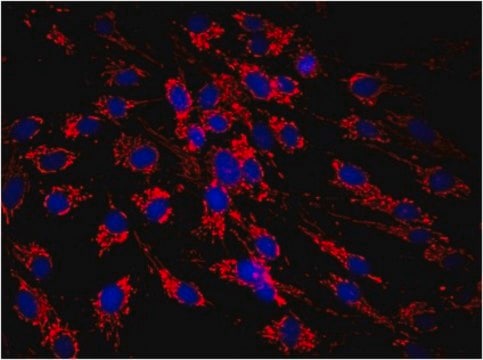
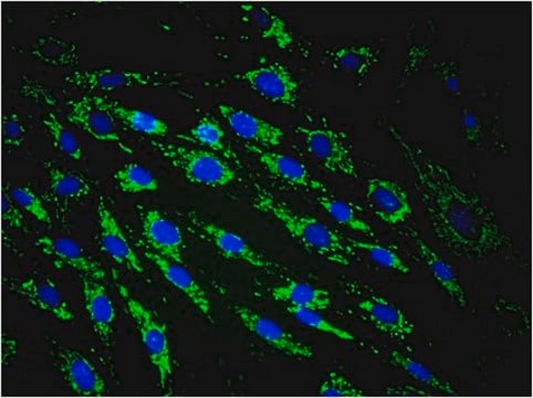
This antibody recognizes a 60 kDa nonglycosylated protein component of mitochondria found in human cells. It can be used to stain mitochondria in cell, tissue or biochemical preparations and can be used as a marker of the mitochondria in subcellular fractions.

Specificity

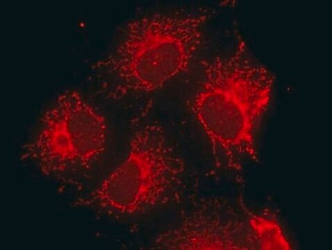
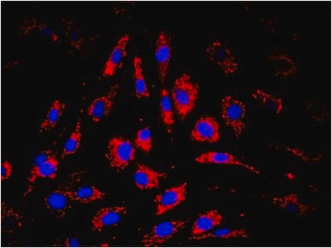
Gives mitochondrial staining on all human cell types and does not cross react with rat and mouse tissue. The staining pattern is best described as a spaghetti-like staining pattern in fibroblasts and a perinuclear large speckled pattern in lymphonoid cells.
MAB1273 recognizes a 65 kD protein by immunoprecipitation. Antibody produces the stringy, "spaghetti-like" staining patten in the cytoplasm of human cells.
Reactivity with other species has not been determined

Immunogen

Epitope: surface of intact mitochondria

Application

Anti-Mitochondria Antibody, surface of intact mitochondria, clone 113-1 is an antibody against Mitochondria for use in IF, IH, IH(P) & IP.
Immunohistochemistry:
1:100 dilutoin from a previous lot was used. Works best with organic fixatives (acetone, methanol, etc). 2% PFA for 10-15 min can be used but stronger use of formalin or 4% PFA limits staining.
The antibody has been used to isolate intact mitochondria from freeze/thaw lysates of living cells via conjugation to magnetic beads {http://www.dynalbiotech.com}.

Immunohistochemistry in paraffin embedded tissues:
Requires citric acid/microwave antigen retrieval; enhanced ABC or amplified detection systems recommended.
Optimal Staining With EDTA Buffer, pH 8.0, Epitope Retrieval: Heart Ventricle Epithelium

The antibody has been used to isolate intact mitochondria from freeze/thaw lysates of living cells via conjugation to magnetic beads {http://www.dynalbiotech.com}.

Indirect immunofluorescence:
1:10-1:50 dilution from a previous lot was used, 30 - 100 μL per slide well.

MAB1273 recognizes a 65 kD protein by immunoprecipitation.

Optimal working dilutions and protocols must be determined by the end user.

Quality

Routinely evaluated by immunohistochemistry on heart ventricle cells.

Immunohistochemistry(paraffin) Analysis:
Mitochondria (cat. # MAB1273) staining on heart ventricle cells. Tissue pretreated with EDTA, pH 8.0This lot of antibody was diluted to 1:80, using IHC-Select Detection with HRP-DAB. Immunoreactivity is seeing the circular-granular layer surrounding the nucleus of the cells.
Optimal Staining With EDTA Buffer, pH 8.0, Epitope Retrieval: Heart Ventricle Epithelium

Target description

65 kDa

Physical form

Format: Purified
Purified mouse monoclonal IgG1 in buffer containing liquid in PBS. Contains no preservative. Concentrated hybridoma supernatant.

Storage and Stability

Stable for 6 months at -20°C in undiluted aliquots from date of receipt.

Analysis Note

Control
COS7 cells.

Other Notes

Concentration: Please refer to the Certificate of Analysis for the lot-specific concentration.

Legal Information

CHEMICON is a registered trademark of Merck KGaA, Darmstadt, Germany

Not finding the right product?  

Try our Product Selector Tool.

Storage Class Code

12 - Non Combustible Liquids

WGK

WGK 2

Flash Point(F)

Not applicable

Flash Point(C)

Not applicable


Certificates of Analysis (COA)

Search for Certificates of Analysis (COA) by entering the products Lot/Batch Number. Lot and Batch Numbers can be found on a product’s label following the words ‘Lot’ or ‘Batch’.

Already Own This Product?

Find documentation for the products that you have recently purchased in the Document Library.

Visit the Document Library

David S Liu et al.
Oncotarget, 7(50), 83342-83358 (2016-11-20)
There is currently a paucity of preclinical models available to study the metastatic process in esophageal cancer. Here we report FLO-1, and its isogenic derivative FLO-1LM, as two spontaneously metastatic cell line models of human esophageal adenocarcinoma. We show that
Sifeng Qu et al.
British journal of cancer, 118(6), 802-812 (2018-01-31)
Docetaxel used for first-line treatment of advanced prostate cancer (PCa) is only marginally effective. We previously showed, using the LTL-313H subrenal capsule patient-derived metastatic PCa xenograft model, that docetaxel combined with Aneustat (OMN54), a multivalent plant-derived therapeutic, led to marked
Jane E Girling et al.
Reproductive biology and endocrinology : RB&E, 8, 84-84 (2010-07-10)
It has been hypothesised that increased VEGF-D expression may be an independent prognostic factor for endometrial cancer progression and lymph node metastasis; however, the mechanism by which VEGF-D may promote disease progression in women with endometrial cancer has not been
Tao Yin et al.
Molecular medicine reports, 12(4), 5093-5099 (2015-08-05)
Ischemic diseases represent a challenging worldwide health burden. The current study investigated the therapeutic potential of genetically modified human placenta‑derived mesenchymal stem cells (hPDMSCs) with basic fibroblast growth factor (FGF2) and platelet‑derived growth factor‑BB (PDGF‑BB) genes in hindlimb ischemia. Mesenchymal
Remedios Castello-Cros et al.
Cell cycle (Georgetown, Tex.), 10(12), 2021-2034 (2011-06-08)
We have previously demonstrated that loss of stromal caveolin-1 (Cav-1) in cancer-associated fibroblasts is a strong and independent predictor of poor clinical outcome in human breast cancer patients. However, the signaling mechanism(s) by which Cav-1 downregulation leads to this tumor-promoting

Our team of scientists has experience in all areas of research including Life Science, Material Science, Chemical Synthesis, Chromatography, Analytical and many others.

Contact Technical Service