Skip to Content
Merck
All Photos(2)

Key Documents

ABE605

Sigma-Aldrich

Anti-TREX1 Antibody

from rabbit, purified by affinity chromatography

Synonym(s):

Three prime repair exonuclease 1, CRV, AGS1, DRN3, HERNS, 3′-5′ exonuclease TREX1, DNase III

Sign Into View Organizational & Contract Pricing


About This Item

UNSPSC Code:
12352203
eCl@ss:
32160702
NACRES:
NA.41
clone:
polyclonal
application:
IF
IHC
WB
species reactivity:
human
technique(s):
immunofluorescence: suitable
immunohistochemistry: suitable
western blot: suitable
citations:

biological source

rabbit

Quality Level

antibody form

affinity isolated antibody

antibody product type

primary antibodies

clone

polyclonal

purified by

affinity chromatography

species reactivity

human

technique(s)

immunofluorescence: suitable
immunohistochemistry: suitable
western blot: suitable

UniProt accession no.

shipped in

wet ice

target post-translational modification

unmodified

Gene Information

human ... TREX1(11277)

General description

TREX1, (DNase III), or three prime repair exonuclease 1 is the major 3’→ 5’ DNA exonuclease in mammalian cells. TREX1 binds preferentially to ssDNA and can also remove mismatched 3’ terminal deoxyribonucleotides at DNA strand breaks. The protein is required for proper DNA checkpoint signaling and plays critical roles in DNA replication and gap filling during DNA repair. TREX1 has a perinuclear distribution in mammalian cells and is colocalized with ER-associated proteins in cells at rest. TREX1 relocalizes to the nucleus and specifically to replication forks during S phase and periods of DNA synthesis and repair such as after irradiation. TREX1 acts on an ssDNA polynucleotide species arising from the processing of aberrant DNA replication intermediates and thus acts to attenuate DNA damage checkpoint signaling and prevent pathological immune activation in normal cells. Defects in TREX1 protein have been associated in humans with many inherited diseases including Aicardi-Goutiėres syndrome type 1 (AGS1), systemic lupus erythematosus (SLE), chilblain lupus type 1 (CHBL1), and vasculopathy, retinal, with cerebral leukodystrophy (RVCL). EMD Millipore’s Anti-TREX1 rabbit polyclonal antibody has been tested in western blotting, immunohistochemistry, and immunofluorescence in human samples.

Immunogen

KLH-conjugated linear peptide corresponding to human TREX1.

Application

Detect DNASE using this rabbit polyclonal antibody, Anti-TREX1 Antibody validated for use in western blotting, IHC & Immunofluorescence.
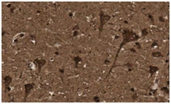
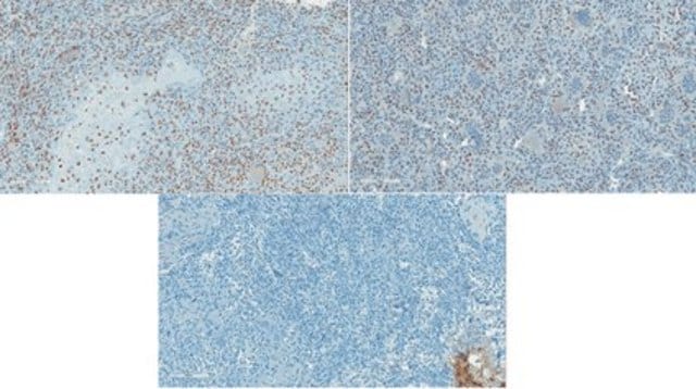
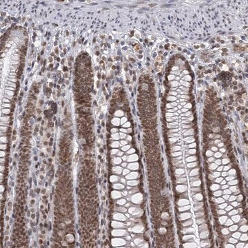
Immunohistochemistry Analysis: 2.5 µg/mL from a representative lot detected TREX1 in human spleen tissue.

Immunofluorescence Analysis: 20 μg/mL from a representative lot detected TREX1 in human spleen tissue.

Quality

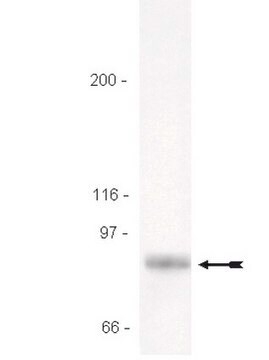
Evaluated by Western Blotting in human spleen tissue lysate.

Western Blotting Analysis: 1 µg/mL of this antibody detected TREX1 in 15 µg of human spleen tissue lysate.

Target description

~30 kDa observed. Uncharacterized bands may appear in some lysate(s). Uniprot describes 3 isoforms at ~32 kDa, ~33 kDa, and ~39 kDa

Other Notes

Concentration: Please refer to the Certificate of Analysis for the lot-specific concentration.

Not finding the right product?  

Try our Product Selector Tool.

Storage Class Code

12 - Non Combustible Liquids

WGK

WGK 2

Flash Point(F)

Not applicable

Flash Point(C)

Not applicable


Certificates of Analysis (COA)

Search for Certificates of Analysis (COA) by entering the products Lot/Batch Number. Lot and Batch Numbers can be found on a product’s label following the words ‘Lot’ or ‘Batch’.

Already Own This Product?

Find documentation for the products that you have recently purchased in the Document Library.

Visit the Document Library

Our team of scientists has experience in all areas of research including Life Science, Material Science, Chemical Synthesis, Chromatography, Analytical and many others.

Contact Technical Service