Saltar al contenido
Merck
Todas las fotos(6)

Documentos

HPA003733

Sigma-Aldrich

Anti-THY1 antibody produced in rabbit

enhanced validation

Prestige Antibodies® Powered by Atlas Antibodies, affinity isolated antibody, buffered aqueous glycerol solution

Sinónimos:

Anti-CD90 antigen, Anti-CDw90, Anti-Thy-1 antigen, Anti-Thy-1 membrane glycoprotein precursor

Iniciar sesiónpara Ver la Fijación de precios por contrato y de la organización


About This Item

MDL number:
UNSPSC Code:
12352203
Human Protein Atlas Number:
NACRES:
NA.41

biological source

rabbit

Quality Level

conjugate

unconjugated

antibody form

affinity isolated antibody

antibody product type

primary antibodies

clone

polyclonal

product line

Prestige Antibodies® Powered by Atlas Antibodies

form

buffered aqueous glycerol solution

species reactivity

human

enhanced validation

orthogonal RNAseq
Learn more about Antibody Enhanced Validation

technique(s)

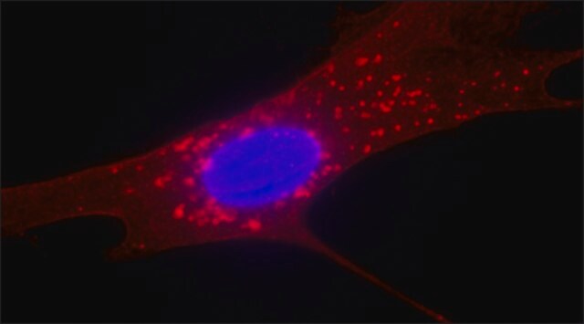
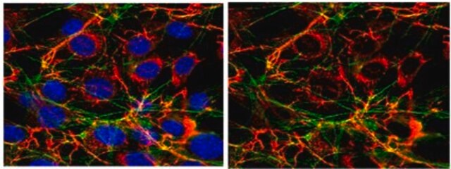
immunofluorescence: 0.25-2 μg/mL
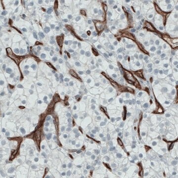
immunohistochemistry: 1:50-1:200

immunogen sequence

VTSLTACLVDQSLRLDCRHENTSSSPIQYEFSLTRETKKHVLFGTVGVPEHTYRSRTNFTSKYNMKVLYLSAFTSKDEGTYTCALHHSGHSPPISSQNVTVLRDKLVKCEGISLLAQNTSWL

UniProt accession no.

shipped in

wet ice

storage temp.

−20°C

target post-translational modification

unmodified

Gene Information

human ... THY1(7070)

General description

Thy-1 (Thy-1 cell surface antigen) is a 18,000Da glycoprotein belonging to the immunoglobulin supergene family. It consists of a hydrophobic segment at the carboxyl terminus and a site for N-glycosylation.
Thy-1 cell surface antigen (Thy-1)/CD90 (cluster of differentiation 90) is a 25–37 kDa glycosylphosphatidylinositol (GPI)-anchored glycoprotein. It was first detected in the T cells of mice. Thy-1 is expressed in thymocytes, T cells, neurons, hematopoietic stem cells, cancer stem cells, endothelial cells and fibroblasts. This gene is located on human chromosome 11q23.

Immunogen

Thy-1 membrane glycoprotein precursor recombinant protein epitope signature tag (PrEST)

Application

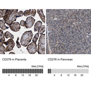
All Prestige Antibodies Powered by Atlas Antibodies are developed and validated by the Human Protein Atlas (HPA) project and as a result, are supported by the most extensive characterization in the industry.

The Human Protein Atlas project can be subdivided into three efforts: Human Tissue Atlas, Cancer Atlas, and Human Cell Atlas. The antibodies that have been generated in support of the Tissue and Cancer Atlas projects have been tested by immunohistochemistry against hundreds of normal and disease tissues and through the recent efforts of the Human Cell Atlas project, many have been characterized by immunofluorescence to map the human proteome not only at the tissue level but now at the subcellular level. These images and the collection of this vast data set can be viewed on the Human Protein Atlas (HPA) site by clicking on the Image Gallery link. We also provide Prestige Antibodies® protocols and other useful information.

Biochem/physiol Actions

Thy-1 cell surface antigen (Thy-1)/CD90 (cluster of differentiation 90) is involved in T-cell activation, neuritis outgrowth modulation, vesicular release of neurotransmitter at the synapse, astrocyte adhesion, apoptosis in carcinoma cells, tumour suppression, wound healing, fibrosis and fibrogenesis. It also controls fibroblast focal adhesion, cytoskeleton organization and cell migration.
Thy-1 is also known as CD90 (Cluster of Differentiation 90). The gene is involved in invasion and associated with epithelial-mesenchymal transition. It is a novel diagnostic marker that contributes for the diagnosis of epithelioid mesothelioma. It can act as a promising novel prognostic marker for male breast cancer. It may act as a biomarker for several tumors as well as cancer stem cells and may be involved in the progression of hepatocellular carcinoma (HCC). It may also act as a potential marker of lung cancer stem cells.

Features and Benefits

Prestige Antibodies® are highly characterized and extensively validated antibodies with the added benefit of all available characterization data for each target being accessible via the Human Protein Atlas portal linked just below the product name at the top of this page. The uniqueness and low cross-reactivity of the Prestige Antibodies® to other proteins are due to a thorough selection of antigen regions, affinity purification, and stringent selection. Prestige antigen controls are available for every corresponding Prestige Antibody and can be found in the linkage section.

Every Prestige Antibody is tested in the following ways:
  • IHC tissue array of 44 normal human tissues and 20 of the most common cancer type tissues.
  • Protein array of 364 human recombinant protein fragments.

Linkage

Corresponding Antigen APREST77497

Physical form

Solution in phosphate-buffered saline, pH 7.2, containing 40% glycerol and 0.02% sodium azide

Legal Information

Prestige Antibodies is a registered trademark of Merck KGaA, Darmstadt, Germany

Disclaimer

Unless otherwise stated in our catalog or other company documentation accompanying the product(s), our products are intended for research use only and are not to be used for any other purpose, which includes but is not limited to, unauthorized commercial uses, in vitro diagnostic uses, ex vivo or in vivo therapeutic uses or any type of consumption or application to humans or animals.

¿No encuentra el producto adecuado?  

Pruebe nuestro Herramienta de selección de productos.

Storage Class

10 - Combustible liquids

wgk_germany

WGK 1

ppe

Eyeshields, Gloves, multi-purpose combination respirator cartridge (US)


Certificados de análisis (COA)

Busque Certificados de análisis (COA) introduciendo el número de lote del producto. Los números de lote se encuentran en la etiqueta del producto después de las palabras «Lot» o «Batch»

¿Ya tiene este producto?

Encuentre la documentación para los productos que ha comprado recientemente en la Biblioteca de documentos.

Visite la Librería de documentos

Xiuping Yan et al.
Oncology reports, 30(6), 2733-2740 (2013-10-09)
Accumulating evidence supports that cancer stem cells (CSCs) are responsible for tumor initiation, progression, distal metastasis and even drug resistance. Although CD90 has been identified as a marker for several types of stem cells, such as liver CSCs, the potential
Kiyoko Kawamura et al.
American journal of clinical pathology, 140(4), 544-549 (2013-09-21)
To pathologically distinguish mesothelioma from lung carcinoma, particularly adenocarcinoma. We conducted immunohistochemical analyses on clinical specimens, including 26 cases of mesothelioma, 28 cases of lung adenocarcinoma, and 33 cases of lung squamous cell carcinoma. We found that CD90 expression was
T Seki et al.
Proceedings of the National Academy of Sciences of the United States of America, 82(19), 6657-6661 (1985-10-01)
The human Thy-1 gene has been isolated and sequenced and compared to the rat and mouse Thy-1 genes. All three genes are organized in the same way: one exon encoding the majority of the signal peptide, another encoding the transmembrane
Caecilia Hapsari Ceriapuri Sukowati et al.
PloS one, 8(10), e76830-e76830 (2013-10-12)
Although the CD90 (Thy-1) was proposed as biomarker of several tumors and cancer stem cells, the involvement of this molecule in the progression of hepatocellular carcinoma (HCC) and other less frequent hepatic neoplasms is still undefined. The distribution of CD90
Ida Johansson et al.
PloS one, 8(10), e78299-e78299 (2013-11-07)
The rapidly growing collection of diverse genome-scale data from multiple tumor types sheds light on various aspects of the underlying tumor biology. With the objective to identify genes of importance for breast tumorigenesis in men and to enable comparisons with

Artículos

Mesenchymal stem cell markers and antibodies suitable for investigating targets in fibroblasts, chondrocytes, adipocytes, osteoblasts, and muscle cells.

Mesenchymal stem cell markers and antibodies suitable for investigating targets in fibroblasts, chondrocytes, adipocytes, osteoblasts, and muscle cells.

Mesenchymal stem cell markers and antibodies suitable for investigating targets in fibroblasts, chondrocytes, adipocytes, osteoblasts, and muscle cells.

Mesenchymal stem cell markers and antibodies suitable for investigating targets in fibroblasts, chondrocytes, adipocytes, osteoblasts, and muscle cells.

Nuestro equipo de científicos tiene experiencia en todas las áreas de investigación: Ciencias de la vida, Ciencia de los materiales, Síntesis química, Cromatografía, Analítica y muchas otras.

Póngase en contacto con el Servicio técnico