
HPA003230
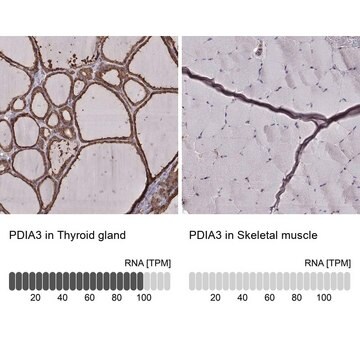
Anti-PDIA3 antibody produced in rabbit
Ab2, Prestige Antibodies® Powered by Atlas Antibodies, affinity isolated antibody, buffered aqueous glycerol solution
Sinónimos:
Anti-58 kDa glucose-regulated protein antibody produced in rabbit, Anti-58 kDa microsomal protein antibody produced in rabbit, Anti-Disulfide isomerase ER-60 antibody produced in rabbit, Anti-ERp57 antibody produced in rabbit, Anti-ERp60 antibody produced in rabbit, Anti-Protein disulfide-isomerase A3 precursor antibody produced in rabbit, Anti-p58 antibody produced in rabbit
About This Item
Productos recomendados


origen biológico
rabbit
Nivel de calidad
conjugado
unconjugated
forma del anticuerpo
affinity isolated antibody
tipo de anticuerpo
primary antibodies
clon
polyclonal
Línea del producto
Prestige Antibodies® Powered by Atlas Antibodies
Formulario
buffered aqueous glycerol solution
reactividad de especies
mouse, rat, human
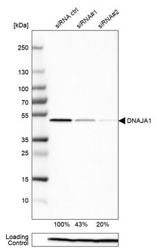
validación mejorada
RNAi knockdown
orthogonal RNAseq
independent
Learn more about Antibody Enhanced Validation
técnicas
immunoblotting: 0.04-0.4 μg/mL
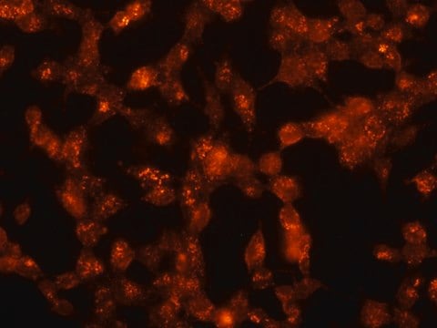
immunofluorescence: 0.25-2 μg/mL
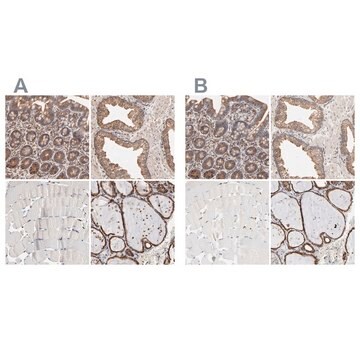
immunohistochemistry: 1:2500-1:5000
secuencia del inmunógeno
PTLKIFRDGEEAGAYDGPRTADGIVSHLKKQAGPASVPLRTEEEFKKFISDKDASIVGFFDDSFSEAHSEFLKAASNLRDNYRFAHTNVESLVNEYDDNGEGIILFRPSHLTNKFEDK
Nº de acceso UniProt
Condiciones de envío
wet ice
temp. de almacenamiento
−20°C
modificación del objetivo postraduccional
unmodified
Información sobre el gen
human ... PDIA3(2923)
¿Está buscando productos similares? Visita Guía de comparación de productos
Descripción general
Inmunógeno
Aplicación
Immunohistochemistry (1 paper)
Western Blotting (1 paper)
Acciones bioquímicas o fisiológicas
Características y beneficios
Every Prestige Antibody is tested in the following ways:
- IHC tissue array of 44 normal human tissues and 20 of the most common cancer type tissues.
- Protein array of 364 human recombinant protein fragments.
Ligadura / enlace
Forma física
Información legal
Cláusula de descargo de responsabilidad
¿No encuentra el producto adecuado?
Pruebe nuestro Herramienta de selección de productos.
Código de clase de almacenamiento
10 - Combustible liquids
Clase de riesgo para el agua (WGK)
WGK 1
Punto de inflamabilidad (°F)
Not applicable
Punto de inflamabilidad (°C)
Not applicable
Equipo de protección personal
Eyeshields, Gloves, multi-purpose combination respirator cartridge (US)
Elija entre una de las versiones más recientes:
Certificados de análisis (COA)
¿No ve la versión correcta?
Si necesita una versión concreta, puede buscar un certificado específico por el número de lote.
¿Ya tiene este producto?
Encuentre la documentación para los productos que ha comprado recientemente en la Biblioteca de documentos.
Nuestro equipo de científicos tiene experiencia en todas las áreas de investigación: Ciencias de la vida, Ciencia de los materiales, Síntesis química, Cromatografía, Analítica y muchas otras.
Póngase en contacto con el Servicio técnico