Saltar al contenido
Merck
Todas las fotos(3)

Documentos clave

MABS1266

Sigma-Aldrich

Anti-LOX-1 Antibody, clone 15C4

clone 15C4, from mouse

Sinónimos:

Oxidized low-density lipoprotein receptor 1, Ox-LDL receptor 1, C-type lectin domain family 8 member A, Lectin-like oxidized LDL receptor 1, Lectin-like oxLDL receptor 1, hLOX-1, Lectin-type oxidized LDL receptor 1

Iniciar sesiónpara Ver la Fijación de precios por contrato y de la organización


About This Item

Código UNSPSC:
12352203
eCl@ss:
32160702
NACRES:
NA.41

origen biológico

mouse

Nivel de calidad

forma del anticuerpo

purified immunoglobulin

tipo de anticuerpo

primary antibodies

clon

15C4, monoclonal

reactividad de especies

human

técnicas

flow cytometry: suitable
immunofluorescence: suitable
immunohistochemistry: suitable
western blot: suitable

isotipo

IgG2aκ

Nº de acceso NCBI

Nº de acceso UniProt

Condiciones de envío

ambient

modificación del objetivo postraduccional

unmodified

Información sobre el gen

human ... OLR1(4973)

Descripción general

Oxidized low-density lipoprotein receptor 1 (UniProt: P78380; also known as Ox-LDL receptor 1, C-type lectin domain family 8 member A, Lectin-like oxidized LDL receptor 1, LOX-1, Lectin-like oxLDL receptor 1, hLOX-1, Lectin-type oxidized LDL receptor 1) is encoded by the OLR1 (also known as CLEC8A, LOX1) gene (Gene ID: 4973) in human. LOX1 is a type II transmembrane receptor for oxidatively modified low density lipoprotein (oxLDL), a well-known marker of atherosclerosis. A free form of LOX-1 has also been reported. LOX-1 has a The C-type lectin domain that mediates the recognition and binding of oxLDL. Its cytoplasmic region is considered to be essential for subcellular sorting on the cell surface. LOX-1 is expressed at high level in endothelial cells and vascular-rich organs such as placenta, lung, liver and brain, aortic intima, bone marrow, spinal cord and substantia nigra. It is also expressed at the surface of dendritic cells. LOX-1 mediates the recognition, internalization and degradation of oxLDL by vascular endothelial cells. OxLDL induces vascular endothelial cell activation and dysfunction, resulting in pro-inflammatory responses, pro-oxidative conditions and also apoptosis. In addition to binding oxLDL, LOX-1 also acts as a receptor for the HSP70 protein involved in antigen cross-presentation to naive T-cells in dendritic cells, thereby participating in cell-mediated antigen cross-presentation. LOX-1 is up-regulated in atherosclerotic lesions, by oxLDL, reactive oxygen species and fluid shear stress.

Especificidad

Clone 15C4 recognizes LOX-1 in human cells.

Inmunógeno

Human LOX 1 ectodomain-hIgGFc fusion protein.

Aplicación

Detect LOX-1 using this mouse monoclonal Anti-LOX-1 Antibody, clone 15C4, Cat. No. MABS1266. It has been tested in Cell Differentiation studies, Flow Cytometry, Immunofluorescence, Immunohistochemistry, and Western Blotting.
Immunohistochemistry Analysis: A 1:250-1,000 dilution from a representative lot detected LOX-1 in human spleen, human large intestine, and human vagina tissues.

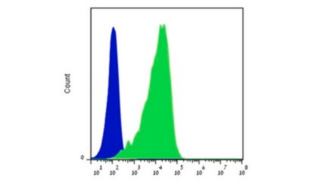
Flow Cytometry Analysis: A representative lot detected LOX-1 in Flow Cytometry applications (Li, D., et. al. (2012). J Exp Med. 209(1):109-21).

Immunofluorescence Analysis: A representative lot detected LOX-1 in Immunofluorescence applications (Li, D., et. al. (2012). J Exp Med. 209(1):109-21).

Cell Differentiation Analysis: A representative lot detected LOX-1 in Cell Differentiation applications (Joo, H., et. al. (2014). Immunity. 41(4):592-604).

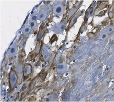
Immunohistochemistry Analysis: A representative lot detected LOX-1 in Immunohistochemistry applications (Duluc, D., et. al. (2013). Microb Pathog. 58:35-44).

Flow Cytometry Analysis: 1 ug from a representative lot detected LOX-1 in one million A549 cells.
Research Category
Signaling

Calidad

Evaluated by Western Blotting in human liver tissue lysates.

Western Blotting Analysis: 2 µg/mL of this antibody detected LOX-1 in 10 µg of human liver tissue lysates.

Descripción de destino

30.96 kDa calculated. Uncharacterized band(s) may appear in some lysates.

Forma física

Format: Purified
Protein G purified
Purified mouse monoclonal antibody IgG2a in PBS without preservatives.

Almacenamiento y estabilidad

Stable for 1 year at -20°C from date of receipt.

Handling Recommendations: Upon receipt and prior to removing the cap, centrifuge the vial and gently mix the solution. Aliquot into microcentrifuge tubes and store at -20°C. Avoid repeated freeze/thaw cycles, which may damage IgG and affect product performance.

Otras notas

Concentration: Please refer to lot specific datasheet.

Cláusula de descargo de responsabilidad

Unless otherwise stated in our catalog or other company documentation accompanying the product(s), our products are intended for research use only and are not to be used for any other purpose, which includes but is not limited to, unauthorized commercial uses, in vitro diagnostic uses, ex vivo or in vivo therapeutic uses or any type of consumption or application to humans or animals.

¿No encuentra el producto adecuado?  

Pruebe nuestro Herramienta de selección de productos.

Código de clase de almacenamiento

12 - Non Combustible Liquids

Clase de riesgo para el agua (WGK)

WGK 2

Punto de inflamabilidad (°F)

Not applicable

Punto de inflamabilidad (°C)

Not applicable


Certificados de análisis (COA)

Busque Certificados de análisis (COA) introduciendo el número de lote del producto. Los números de lote se encuentran en la etiqueta del producto después de las palabras «Lot» o «Batch»

¿Ya tiene este producto?

Encuentre la documentación para los productos que ha comprado recientemente en la Biblioteca de documentos.

Visite la Librería de documentos

Nuestro equipo de científicos tiene experiencia en todas las áreas de investigación: Ciencias de la vida, Ciencia de los materiales, Síntesis química, Cromatografía, Analítica y muchas otras.

Póngase en contacto con el Servicio técnico