Saltar al contenido
Merck
Todas las fotos(3)

Key Documents

MABN125

Sigma-Aldrich

Anti-DRD4 Antibody, clone 2B9

clone 2B9, from mouse

Sinónimos:

D(4) dopamine receptor, D(2C) dopamine receptor, Dopamine D4 receptor

Iniciar sesiónpara Ver la Fijación de precios por contrato y de la organización


About This Item

UNSPSC Code:
12352203
eCl@ss:
32160702
NACRES:
NA.41

biological source

mouse

Quality Level

antibody form

purified immunoglobulin

antibody product type

primary antibodies

clone

2B9, monoclonal

species reactivity

human, rat, mouse

technique(s)

immunocytochemistry: suitable
western blot: suitable

isotype

IgG1κ

NCBI accession no.

UniProt accession no.

shipped in

wet ice

target post-translational modification

unmodified

Gene Information

mouse ... Drd4(13491)

General description

Five subtypes of dopamine receptors exist in two subfamilies: two D1-like (D1 and D5) and three D2-like (D2, D3 and D4). As members of the G protein-coupled receptor (GPCR) family, dopamine receptors initiate their biological function by coupling to GTP regulatory proteins (G proteins). D1-like receptors, DRD1 and DRD5, stimulate adenylyl cyclase whereas D2-like receptors, DRD2, DRD3 and DRD4, are coupled to inhibitory G proteins and their stimulation leads to the inhibition of adenylyl cyclase activity, and subsequent respective cellular signaling. All five subtypes have seven transmembrane domains, an extracellular amino terminus, and an intracellular carboxyl terminus. Allelic variants of the D4 dopamine receptor (DRD4) are known characterized risk factors of ADHD. It is reported that hypoxia enhances DRD4 promoter activity by several fold. It is also thought that dopamine receptors such as DRD4 are involved in neuroexcitation and in a variety of other neurological and psychiatric disorders.

Immunogen

Linear peptide corresponding to human DRD4.

Application

Research Category
Neuroscience
Research Sub Category
Neurotransmitters & Receptors
This Anti-DRD4 Antibody, clone 2B9 is validated for use in WB, IC for the detection of DRD4.
Western Blot Analysis: 0.5 µg/mL from a representative lot detected DRD4 in 10µg of rat brain tissue lysate.

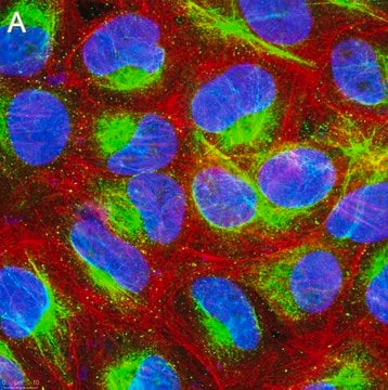
Immunocytochemistry Analysis: A representative lot from an independent laboratory detected DRD4 in Ntera-2 cells and in rat brain neurons. (Wolstencroft, E.C., et al. (2007). J Mol Histol. 38(4):333-340.)

Quality

Evaluated by Western Blot in mouse brain tissue lysate.

Western Blot Analysis: 0.5 µg/mL of this antibody detected DRD4 in 10µg of mouse brain tissue lysate.

Target description

~ 48 kDa observed.

Physical form

Format: Purified
Protein G Purified
Purified mouse monoclonal IgG1κ in buffer containing 0.1 M Tris-Glycine (pH 7.4), 150 mM NaCl with 0.05% sodium azide.

Storage and Stability

Stable for 1 year at 2-8°C from date of receipt.

Analysis Note

Control
Mouse brain tissue lysate.

Other Notes

Concentration: Please refer to the Certificate of Analysis for the lot-specific concentration.

Disclaimer

Unless otherwise stated in our catalog or other company documentation accompanying the product(s), our products are intended for research use only and are not to be used for any other purpose, which includes but is not limited to, unauthorized commercial uses, in vitro diagnostic uses, ex vivo or in vivo therapeutic uses or any type of consumption or application to humans or animals.

¿No encuentra el producto adecuado?  

Pruebe nuestro Herramienta de selección de productos.

Storage Class

12 - Non Combustible Liquids

wgk_germany

WGK 1

flash_point_f

Not applicable

flash_point_c

Not applicable


Certificados de análisis (COA)

Busque Certificados de análisis (COA) introduciendo el número de lote del producto. Los números de lote se encuentran en la etiqueta del producto después de las palabras «Lot» o «Batch»

¿Ya tiene este producto?

Encuentre la documentación para los productos que ha comprado recientemente en la Biblioteca de documentos.

Visite la Librería de documentos

Sohn G Kim et al.
Journal of leukocyte biology, 94(4), 835-843 (2013-07-03)
In vivo and in vitro exposure to stimulants has been associated with increased levels of HIV infection in PBMCs. Among these lymphocyte subsets, quiescent CD4(+) T cells make up the majority of circulating T cells in the blood. Others and
Leigh Dairaghi et al.
eNeuro, 9(2) (2022-02-16)
Gonadotropin-releasing hormone (GnRH)-secreting neurons control fertility. The release of GnRH peptide regulates the synthesis and release of both luteinizing hormone (LH) and Follicle stimulation hormone (FSH) from the anterior pituitary. While it is known that dopamine regulates GnRH neurons, the
Wei Wang et al.
Immunity, 51(6), 1102-1118 (2019-11-24)
Young children are more susceptible to developing allergic asthma than adults. As neural innervation of the peripheral tissue continues to develop after birth, neurons may modulate tissue inflammation in an age-related manner. Here we showed that sympathetic nerves underwent a

Nuestro equipo de científicos tiene experiencia en todas las áreas de investigación: Ciencias de la vida, Ciencia de los materiales, Síntesis química, Cromatografía, Analítica y muchas otras.

Póngase en contacto con el Servicio técnico