Saltar al contenido
Merck
Todas las fotos(2)

Key Documents

07-403-I

Sigma-Aldrich

Anti-Cu/Zn-SOD Antibody

from rabbit, purified by affinity chromatography

Sinónimos:

Superoxide dismutase [Cu-Zn], Cu/Zn-SOD, Superoxide dismutase 1, hSod1

Iniciar sesiónpara Ver la Fijación de precios por contrato y de la organización


About This Item

UNSPSC Code:
12352203
eCl@ss:
32160702
NACRES:
NA.41

biological source

rabbit

Quality Level

antibody form

affinity isolated antibody

antibody product type

primary antibodies

clone

polyclonal

purified by

affinity chromatography

species reactivity

human, mouse, rat

species reactivity (predicted by homology)

monkey (based on 100% sequence homology), primate (based on 100% sequence homology)

technique(s)

immunohistochemistry: suitable
western blot: suitable

NCBI accession no.

UniProt accession no.

shipped in

wet ice

target post-translational modification

unmodified

Gene Information

human ... SOD1(6647)

General description

Superoxide dismutase [Cu-Zn] (EC 1.15.1.1; UniProt P00441; also known as Cu/Zn superoxide dismutase, Epididymis secretory protein Li 44, hSod1, Indophenoloxidase A, Superoxide dismutase 1) is encoded by the SOD1 (also known as ALS1) gene (Gene ID 6647) in human. Superoxide dismutases (SOD) provide important cellular antioxidant defense against oxidative damage by catalyzing the dismutation (or partitioning) of the superoxide (O2−) radical into either molecular oxygen (O2) or hydrogen peroxide (H2O2). Superoxide dismutases are divided into three families based on the metal cofactor. The Cu/Zn type binds both copper and zinc, the Fe and Mn types bind either iron or manganese, and the Ni type binds nickel. Three forms of SOD are present in mammals, the cytosolic SOD1, the mitochondrial SOD2, and the extracellular SOD3. SOD1 exists in dimeric form, whereas the SOD2 and SOD3 are tetrameric. SOD1 and SOD3 Cu/Zn type, whereas SOD2 is Mn type. Mutations in SOD1 gene have been linked to 20% of amyotrophic lateral sclerosis (ALS), also known as Lou Gehrig disease.

Immunogen

Epitope: C-terminal region
KLH-conjugated linear peptide corresponding to a C-terminal sequence of human Cu/Zn-SOD.

Application

Anti-Cu/Zn-SOD Antibody is an antibody against Cu/Zn-SOD for use in Western Blotting, Immunohistochemistry.
Immunohistochemistry Analysis: A 1:1,000 dilution from a representative lot detected Cu/Zn-SOD in human cerebral cortex, mouse cerebellum, and mouse kidney tissue.
Research Category
Neuroscience
Research Sub Category
Developmental Neuroscience

Quality

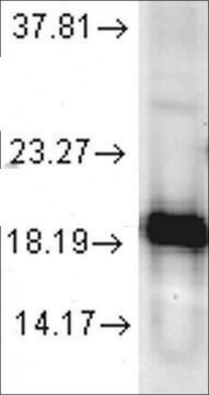
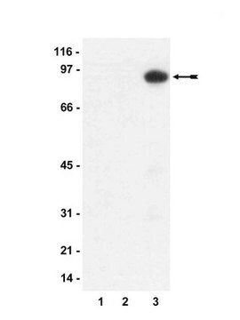
Evaluated by Western Blotting in human brain tissue lysate.

Western Blotting Analysis: 0.1 µg/mL of this antibody detected Cu/Zn-SOD in 10 µg of human brain tissue lysate.

Target description

~16 kDa observed

Physical form

Affinity purified
Purified rabbit polyclonal antibody in buffer containing 0.1 M Tris-Glycine (pH 7.4), 150 mM NaCl with 0.05% sodium azide.

Storage and Stability

Stable for 1 year at 2-8°C from date of receipt.

Other Notes

Concentration: Please refer to lot specific datasheet.

Disclaimer

Unless otherwise stated in our catalog or other company documentation accompanying the product(s), our products are intended for research use only and are not to be used for any other purpose, which includes but is not limited to, unauthorized commercial uses, in vitro diagnostic uses, ex vivo or in vivo therapeutic uses or any type of consumption or application to humans or animals.

¿No encuentra el producto adecuado?  

Pruebe nuestro Herramienta de selección de productos.

Storage Class

12 - Non Combustible Liquids

wgk_germany

WGK 1

flash_point_f

Not applicable

flash_point_c

Not applicable


Certificados de análisis (COA)

Busque Certificados de análisis (COA) introduciendo el número de lote del producto. Los números de lote se encuentran en la etiqueta del producto después de las palabras «Lot» o «Batch»

¿Ya tiene este producto?

Encuentre la documentación para los productos que ha comprado recientemente en la Biblioteca de documentos.

Visite la Librería de documentos

Aisha Al-Kouh et al.
Journal of vascular research, 61(4), 179-196 (2024-07-02)
The comorbidities of ischemic heart disease (IHD) and diabetes mellitus (DM) compromise the protection of the diabetic heart from ischemia/reperfusion (I/R) injury. We hypothesized that manipulation of reperfusion injury salvage kinase (RISK) and survivor activating factor enhancement (SAFE) pathways might
Anne-Sophie Montero et al.
EBioMedicine, 106, 105235-105235 (2024-07-13)
Amyotrophic lateral sclerosis (ALS) is a fatal neurodegenerative disease characterized by a progressive loss of motor neurons. The limited efficacy of recent therapies in clinical development may be linked to lack of drug penetration to the affected motor neurons due
Jyotsana Pandey et al.
The Journal of biological chemistry, 299(5), 104695-104695 (2023-04-13)
Pulmonary fibrosis is a progressive lung disease characterized by macrophage activation. Asbestos-induced expression of nicotinamide adenine dinucleotide phosphate hydrogen oxidase 4 (NOX4) in lung macrophages mediates fibrotic progression by the generation of mitochondrial reactive oxygen species (ROS), modulating mitochondrial biogenesis
Aisha Al-Kouh et al.
Pharmaceuticals (Basel, Switzerland), 16(2) (2023-06-01)
Diabetes mellitus (DM) is a risk factor for cardiovascular diseases, specifically, the ischemic heart diseases (IHD). The renin-angiotensin system (RAS) affects the heart directly and indirectly. However, its role in the protection of the heart against I/R injury is not
Chao He et al.
JCI insight, 4(16) (2019-08-23)
Macrophage activation is implicated in the development of pulmonary fibrosis by generation of profibrotic molecules. Although NADPH oxidase 4 (NOX4) is known to contribute to pulmonary fibrosis, its effects on macrophage activation and mitochondrial redox signaling are unclear. Here, we

Nuestro equipo de científicos tiene experiencia en todas las áreas de investigación: Ciencias de la vida, Ciencia de los materiales, Síntesis química, Cromatografía, Analítica y muchas otras.

Póngase en contacto con el Servicio técnico