Skip to Content
Merck
All Photos(1)

Key Documents

F2772

Sigma-Aldrich

Anti-Mouse IgG (Fc specific) F(ab′)2 fragment−FITC antibody produced in goat

flow cytometry grade, affinity isolated antibody, buffered aqueous solution

Sign Into View Organizational & Contract Pricing


About This Item

MDL number:
UNSPSC Code:
12352203
NACRES:
NA.46

biological source

goat

Quality Level

conjugate

FITC conjugate

antibody form

affinity isolated antibody

antibody product type

secondary antibodies

grade

flow cytometry grade

clone

polyclonal

form

buffered aqueous solution

storage condition

protect from light

technique(s)

flow cytometry: 1:100
immunohistochemistry (formalin-fixed, paraffin-embedded sections): 1:160

storage temp.

−20°C

target post-translational modification

unmodified

Looking for similar products? Visit Product Comparison Guide

General description

IgG antibody subtype is the most abundant serum immunoglobulins of the immune system. It is secreted by B cells and is found in blood and extracellular fluids.
Fluorescein isothiocyanate (FITC) is a fluorescein derivative (fluorochrome) used to tag antibodies, including secondary antibodies, for use in fluorescence-based assays and procedures. FITC excites at 495 nm and emits at 521 nm.
Mouse IgG is a plasma B cell derived antibody isotype defined by its heavy chain. IgG is the most abundant antibody isotype found in mouse serum. IgG crosses the placental barrier, is a complement activator and binds to the Fc-receptors on phagocytic cells. The level of IgG may vary with the status of disease or infection.
Anti-mouse IgG is conjugated to Fluorescein Isothiocyanate (FITC), Isomer I. The F(ab′)2 antibodies isolated from goat antiserum by affinity chromatography, react specifically with mouse IgG Fc; does not bind other mouse Igs. No cross reaction is observed with mouse Fab fragment. The antibody shows minimal cross reaction with human, horse and bovine proteins on tissue or cell preparations.

Specificity

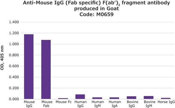
Goat polyclonal anti-Mouse IgG (Fc specific) F(ab′)2 fragment−FITC antibody isolated from goat antiserum by affinity chromatography reacts specifically with mouse Fc fragment. No cross reaction is observed with mouse Fab fragment. The antibodies are adsorbed to assure minimal cross reaction with human, horse and bovine proteins on tissue or cell preparations.

Immunogen

Purified Fc fragments of mouse IgG

Application

Anti-Mouse IgG (Fc specific) F(ab′)2 fragment-FITC antibody produced in goat has been used in :
  • flow cytometry for labelling of human peripheral blood lymphocytes (dilution of 1:100) and label human polymorphonuclear cells (PMNs) (dilution of 1:50).
  • immunocytochemistry using human embryonic stem cells
  • immunohistochemistry at a working dilution of 1:160.
Goat polyclonal anti-Mouse IgG (Fc specific) F(ab′)2 fragment-FITC antibody may be used as a reagent in flow cytometry, immunohistology and immunocytology offering sensitive and specific activity to mouse IgG and no cross reactivity with human, horse or bovine immunoglobulins.
Immunohistology: A minimum dilution of 1:160 is determined by indirect immunofluorescence on formalinfixed, and paraffin-embedded human tonsil using mouse monoclonal anti-human IgG as the primary antibody.

Biochem/physiol Actions

immunoglobulin G (IgG) antibody provides protection from infections caused by bacteria, fungi and viruses. Maternal IgG is transferred to fetus through the placenta that is vital for immune defence of the neonate against infections. IgG antibody participates in complement fixation and opsonization.

Other Notes

Antibody adsorbed with bovine, equine and human serum proteins.

Physical form

Solution in 0.01 M phosphate buffered saline, pH 7.4, containing 1% bovine serum albumin and 15 mM sodium azide.

Preparation Note

Adsorbed to reduce background with bovine, horse or human samples.
Useful when trying to avoid background staining due to the presence of Fc receptors.

Disclaimer

Unless otherwise stated in our catalog or other company documentation accompanying the product(s), our products are intended for research use only and are not to be used for any other purpose, which includes but is not limited to, unauthorized commercial uses, in vitro diagnostic uses, ex vivo or in vivo therapeutic uses or any type of consumption or application to humans or animals.

Not finding the right product?  

Try our Product Selector Tool.

Storage Class Code

10 - Combustible liquids

WGK

nwg

Flash Point(F)

Not applicable

Flash Point(C)

Not applicable

Personal Protective Equipment

dust mask type N95 (US), Eyeshields, Gloves

Certificates of Analysis (COA)

Search for Certificates of Analysis (COA) by entering the products Lot/Batch Number. Lot and Batch Numbers can be found on a product’s label following the words ‘Lot’ or ‘Batch’.

Already Own This Product?

Find documentation for the products that you have recently purchased in the Document Library.

Visit the Document Library

Customers Also Viewed

Pernille B Jørgensen et al.
PloS one, 7(9), e46120-e46120 (2012-10-11)
Multiple sclerosis (MS) is associated with Epstein-Barr virus (EBV) infection, but impaired immune suppression may be part of the disease pathogenesis. CD8(+) T cells that are restricted by HLA-E exert an important immunoregulatory mechanism. To explore how EBV might interfere
Ana Cecilia Machado Diaz et al.
Arthritis, 2012, 943156-943156 (2012-08-14)
Rheumatoid arthritis (RA) is an autoimmune and inflammatory disease in which many cytokines have been implicated. In particular, IL-15 is a cytokine involved in the inflammatory processes and bone loss. The aim of this study was to investigate the existence
Mami Nakahara et al.
Endocrinology, 153(4), 2034-2042 (2012-02-16)
We have recently shown that wild type mice are highly tolerant, whereas thyrotropin receptor (TSHR) knockout (KO) mice are susceptible to immunization with the mouse TSHR, the autoantigen in Graves' disease. However, because TSHR KO mice lack the endogenous TSHR
Thomas Hills et al.
PloS one, 11(11), e0166383-e0166383 (2016-11-20)
The need for CD4+ T cell responses to arise de novo following vaccination can limit the speed of B cell responses. Populations of pre-existing vaccine-induced or anti-viral CD4+ T cells recognising distinct antigens could be exploited to overcome this limitation.
Opsonophagocytosis assay using flow-cytometry.
Joyce S Plested et al.
Methods in molecular medicine, 71, 263-275 (2002-10-11)

Our team of scientists has experience in all areas of research including Life Science, Material Science, Chemical Synthesis, Chromatography, Analytical and many others.

Contact Technical Service