Skip to Content
Merck
All Photos(1)

Key Documents

A7549

Sigma-Aldrich

Anti-Apoptosis-Inducing Factor antibody produced in rabbit

IgG fraction of antiserum, buffered aqueous solution

Synonym(s):

Anti-AIF

Sign Into View Organizational & Contract Pricing


About This Item

MDL number:
UNSPSC Code:
12352203
NACRES:
NA.41

biological source

rabbit

Quality Level

conjugate

unconjugated

antibody form

IgG fraction of antiserum

antibody product type

primary antibodies

clone

polyclonal

form

buffered aqueous solution

mol wt

antigen 57 kDa

species reactivity

mouse, human

packaging

antibody small pack of 25 μL

technique(s)

microarray: suitable
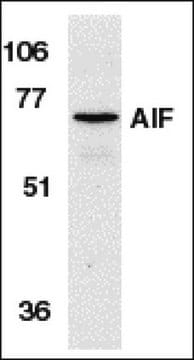
western blot: 1:1,000 using human epitheloid carcinoma HeLa cell extract and mouse brain extract

UniProt accession no.

shipped in

dry ice

storage temp.

−20°C

target post-translational modification

unmodified

Gene Information

human ... AIFM1(9131)
mouse ... Aifm1(26926)

General description

Apoptosis-Inducing Factor (AIF) is a mitochondrial flavoprotein that can induce apoptosis in isolated nuclei. Studies have reported that AIF can induce the realease of cytochrome c and caspase 9. Bcl-2 is known to inhibit AIF release without affecting its apoptotic functions.
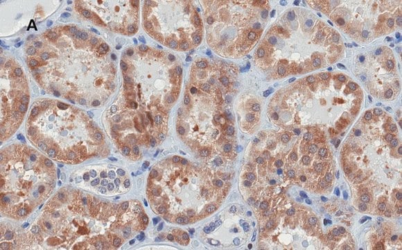
Rabbit Anti-AIF antibody recognizes human and mouse AIF (57kDa). Staining of AIF in immunoblotting is specifically inhibited with the AIF immunizing peptide (human, amino acids 593-613).
Apoptosis-inducing factor (AIF) is encoded by the gene mapped to human chromosome X (Xq25-Xq26). It is a 57 kDa mitochondrial flavoprotein. AIF contains two mitochondrial localization sequences and two putative nuclear localization sequences.

Immunogen

synthetic peptide corresponding to the C-terminus of human Apoptosis Inducing Factor (AIF), (amino acids 593-613), conjugated to KLH.

Application

Protein lysates from 2CLL, 10 MCl and Jeko-1 cells were analyzed by western blot using anti-AIF as the primary antibody. Cells also underwent intracellular delivery by being transfected with anti-AIF at a final dilution of 1:2500 and then analyzed by FLOW cytometry.
Rabbit Anti-Apoptosis-Inducing Factor (AIF) antibody has been used for immunoblot analysis at 1:1000 and 1:2000 dilutions. The antibody can also be used for microarray applications.

Biochem/physiol Actions

Microinjection of apoptosis-inducing factor (AIF) into the cytoplasm of intact cells induces chromatin condensation, dissipation of the mitochondrial transmembrane potential and exposure of phosphatidylserine in the plasma membrane.

Physical form

Solution in 0.01 M phosphate buffered saline, pH 7.4, containing 15 mM sodium azide.

Disclaimer

Unless otherwise stated in our catalog or other company documentation accompanying the product(s), our products are intended for research use only and are not to be used for any other purpose, which includes but is not limited to, unauthorized commercial uses, in vitro diagnostic uses, ex vivo or in vivo therapeutic uses or any type of consumption or application to humans or animals.

Not finding the right product?  

Try our Product Selector Tool.

Storage Class Code

12 - Non Combustible Liquids

WGK

WGK 2

Flash Point(F)

Not applicable

Flash Point(C)

Not applicable


Certificates of Analysis (COA)

Search for Certificates of Analysis (COA) by entering the products Lot/Batch Number. Lot and Batch Numbers can be found on a product’s label following the words ‘Lot’ or ‘Batch’.

Already Own This Product?

Find documentation for the products that you have recently purchased in the Document Library.

Visit the Document Library

Life with or without AIF
Hangen E, et al.
Trends in Biochemical Sciences, 35(5), 278-287 (2010)
Export of mitochondrial AIF in response to proapoptotic stimuli depends on processing at the intermembrane space
Otera H, et al.
The Embo Journal, 24(7), 1375-1386 (2005)
Toxic proteins released from mitochondria in cell death
Saelens X, et al.
Oncogene, 23(16), 2861-2861 (2004)
Virgilia Sahiri et al.
International journal of molecular sciences, 24(3) (2023-02-12)
Focal segmental glomerulosclerosis (FSGS) is a major cause of end-stage renal disease and remains without specific treatment. To identify new events during FSGS progression, we used an experimental model of FSGS associated with nephroangiosclerosis in rats injected with L-NAME (Nω-nitro-L-arginine
Gaël Roué et al.
Clinical cancer research : an official journal of the American Association for Cancer Research, 14(21), 6907-6915 (2008-11-05)
Chronic lymphocytic leukemia (CLL) and mantle cell lymphoma (MCL) are two incurable B-cell lymphoid neoplasms characterized by distinct clinical presentation and evolution. Bendamustine hydrochloride is a multifunctional, alkylating agent with a purine-like ring system that exhibits activity in multiple cancer

Our team of scientists has experience in all areas of research including Life Science, Material Science, Chemical Synthesis, Chromatography, Analytical and many others.

Contact Technical Service