Skip to Content
Merck
All Photos(1)

Key Documents

283M-1

Sigma-Aldrich

HBME-1 (HBME-1) Mouse Monoclonal Antibody

Sign Into View Organizational & Contract Pricing


About This Item

UNSPSC Code:
12352203
NACRES:
NA.41

biological source

mouse

Quality Level

100
500

conjugate

unconjugated

antibody form

culture supernatant

antibody product type

primary antibodies

clone

HBME-1, monoclonal

description

For In Vitro Diagnostic Use in Select Regions (See Chart)

form

buffered aqueous solution

species reactivity

human

packaging

vial of 0.1 mL concentrate (283M-14)
vial of 0.5 mL concentrate (283M-15)
bottle of 1.0 mL predilute (283M-17)
vial of 1.0 mL concentrate (283M-16)
bottle of 7.0 mL predilute (283M-18)

manufacturer/tradename

Cell Marque

technique(s)

immunohistochemistry (formalin-fixed, paraffin-embedded sections): 1:25-1:100

isotype

IgMκ

control

mesothelioma

shipped in

wet ice

storage temp.

2-8°C

visualization

cytoplasmic, membranous

General description

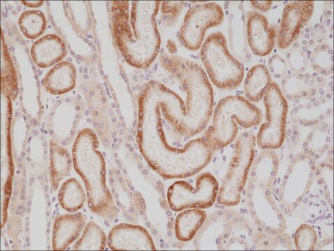
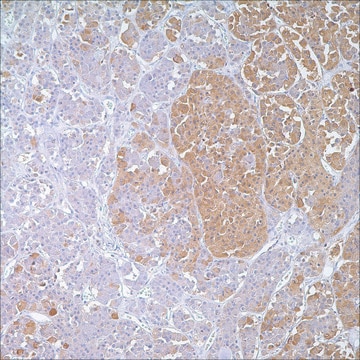
Hector Battifora mesothelial-1 (HBME-1) is a membrane antigen that exists in the microvilli ofmesothelial cells and other epithelial cells. Anti-HBME-1 labels thyroid papillary carcinomaand follicular carcinoma but not normal thyroid making it a valuable marker for distinguishingthyroid malignancies from benign thyroid lesions. It has also been demonstrated to labelmesothelial cells, both benign and malignant (malignant mesothelioma), and thus can aid inthe identification of mesothelioma.

Quality


IVD

IVD

IVD

RUO

Linkage

HBME-1 Positive Control Slides, Product No. 283S, are available for immunohistochemistry (formalin-fixed, paraffin-embedded sections).

Physical form

Solution in Tris Buffer, pH 7.3-7.7, with 1% BSA and <0.1% Sodium Azide

Preparation Note

Download the IFU specific to your product lot and formatNote: This requires a keycode which can be found on your packaging or product label.

Other Notes

For Technical Service please contact: 800-665-7284 or email: service@cellmarque.com

Legal Information

Cell Marque is a trademark of Merck KGaA, Darmstadt, Germany

Not finding the right product?  

Try our Product Selector Tool.


Certificates of Analysis (COA)

Search for Certificates of Analysis (COA) by entering the products Lot/Batch Number. Lot and Batch Numbers can be found on a product’s label following the words ‘Lot’ or ‘Batch’.

Already Own This Product?

Find documentation for the products that you have recently purchased in the Document Library.

Visit the Document Library

A C Bateman et al.
Histopathology, 30(1), 49-56 (1997-01-01)
Histological diagnosis of malignant mesothelioma and differentiation from adenocarcinoma is often difficult. Definitive pathological confirmation of malignant mesothelioma requires demonstration of an appropriate immunohistochemical phenotype. Selection of an optimum panel of immunohistochemical antibodies for the reliable identification of malignant mesothelioma
C C Cheung et al.
Modern pathology : an official journal of the United States and Canadian Academy of Pathology, Inc, 14(4), 338-342 (2001-04-13)
In thyroid, the diagnosis of papillary carcinoma (PC) is based on nuclear features; however, identification of these features is inconsistent and controversial. Proposed markers of PC include HBME-1, specific cytokeratins (CK) such as CK19, and ret, the latter reflecting a
A Coli et al.
Journal of experimental & clinical cancer research : CR, 26(2), 221-227 (2007-08-30)
Ninety-six thyroid lesions were immunohistochemically evaluated for HBME-1 and Galectin-3 expression including nodules with cytological atypia, the latter defined as nuclear features suggestive but not diagnostic of papillary thyroid carcinoma. Thirty nodules with cytological atypia, 49 papillary thyroid carcinomas (PTCs)
Daniela Cabibi et al.
Thyroid : official journal of the American Thyroid Association, 17(7), 603-607 (2007-08-19)
To verify whether immunohistochemistry might be useful in the distinction between a true laterocervical metastasis of an undetected thyroid carcinoma and a primary tumor outside the gland. Galectin-3, cytokeratin 19, and HBME-1 were assessed in six cases (group A) of
J E Barroeta et al.
Endocrine pathology, 17(3), 225-234 (2007-02-20)
Several immunohistochemical markers have been used to aid in the diagnosis of follicular-derived lesions of the thyroid (FDLT). In this study we analyze the diagnostic efficacy of an immunopanel of antibodies to cytokeratin-19 (CK19), galectin-3 (GAL-3), HBME-1, anti-MAP kinase (ERK)

Our team of scientists has experience in all areas of research including Life Science, Material Science, Chemical Synthesis, Chromatography, Analytical and many others.

Contact Technical Service