Skip to Content
Merck
All Photos(4)

Key Documents

MAB3067

Sigma-Aldrich

Anti-Connexin 43 Antibody

CHEMICON®, mouse monoclonal, 4E6.2

Synonym(s):

Gap junction 43 kDa heart protein, connexin 43, gap junction protein, alpha 1, 43kDa, gap junction protein, alpha 1, 43kDa (connexin 43), gap junction protein, alpha-like

Sign Into View Organizational & Contract Pricing


About This Item

UNSPSC Code:
12352203
eCl@ss:
32160702
NACRES:
NA.41

product name

Anti-Connexin 43 Antibody, clone 4E6.2, clone 4E6.2, Chemicon®, from mouse

biological source

mouse

Quality Level

antibody form

purified immunoglobulin

antibody product type

primary antibodies

clone

4E6.2, monoclonal

species reactivity

mouse, rat, pig, canine

species reactivity (predicted by homology)

human

manufacturer/tradename

Chemicon®

technique(s)

ELISA: suitable
immunocytochemistry: suitable
immunohistochemistry: suitable
western blot: suitable

isotype

IgG1

NCBI accession no.

UniProt accession no.

shipped in

wet ice

target post-translational modification

unmodified

Gene Information

human ... GJA1(2697)

General description

Connexins are highly homologous proteins that assemble as hexamers (connexons) to form gap junctions, small channels which allow for the passage of ions and other small molecules up to 1,200 daltons between neighboring cells. Gap junctions play an important role in a variety of cellular processes including homeostasis, intercellular metabolic cooperation and synchronization, morphogenesis, cell differentiation, and growth control. The most broadly studied connexins are the 26, 32, 43 and 45 kDa isoforms. Connexin 43 is a 382 a.a. protein belonging to the alpha-type (Group II) subfamily of connexin proteins and possessing four transmembrane regions, with cytoplasmic amino and carboxyl terminals. It undergoes rapid turnover in the cell and its monomers may reside in the ER/Golgi network, forming a reservoir available for assembly upon degradation of existing connexin-43 channels. Connexin 43 has been detected in a variety of tissues including: atrial and ventricular myocytes, granulose cells, astrocytes, fibroblasts, leptomeningeal cells, brain, lung, smooth muscle, uterine muscle, kidney, lens, epithelium, cornea and testis. Connexin 43 also may have a role in communication between migrating cardiac neural crest cells.

Specificity

Connexin-43. Recognizes proteins of molecular weight 43-47 kDa on Western blots of mouse brain lysate. Mouse connexin 43 sequence is identical in rat and bovine, and differs by one amino acid in pig. This sequence has no known homology with other connexins. Also effective for detection of Cx43 in mouse keratinocyte cells and on frozen sections of rat heart.

Immunogen

A synthetic peptide corresponding to positions 252-270 of the mouse connexin 43 sequence. This sequence is identical in rat and bovine.
Epitope: C-terminus

Application

Anti-Connexin 43 Antibody, clone 4E6.2 detects level of Connexin 43 & has been published & validated for use in ELISA, IC, IH & WB.
Research Category
Cell Structure
Research Sub Category
Adhesion (CAMs)
Western Blot (colorimetric detection):
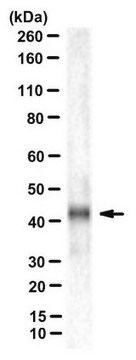
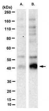
1:1000-10,000 freshly prepared lysates, proteinase inhibitors are recommended. Recognizes proteins of molecular weight 43-47 kDa on Western blots of mouse brain lysate.

Immunocytochemistry:
A 1:50-1:250 dilution of a previous lot was used in immunocytochemistry.

Immunohistochemistry:
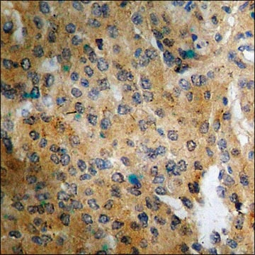
A previous lot was used to detect Connexin 43 expression in the keratinocytes of the middle and upper epidermal layers of frozen human skin tissue (Arita, Ken, et al. (2006). Am. J. Pathol. 169:416-423).

Immunohistochemistry:
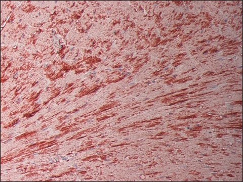
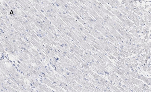
A previous lot was used to detect Connexin 43 expression in human and rat heart tissue (1:500 dilution).

ELISA:
A previous lot of this antibody was used in ELISA.

Optimal working dilutions must be determined by end user.

Quality

Evaluated by Western Blot on rat brain lysates.

Western Blot Analysis:
1:1000 dilution of this antibody detected Connexin 43 on 10 μg of rat brain lysates

Target description

43-47 kDa

Physical form

Format: Purified
Protein A purified
Purified mouse monoclonal IgG1 liquid in PBS containing sodium azide.

Storage and Stability

Stable for 1 year at 2-8ºC in undiluted aliquots from date of receipt.

Analysis Note

Control
Mouse brain tissue lysate

Other Notes

Concentration: Please refer to the Certificate of Analysis for the lot-specific concentration.

Legal Information

CHEMICON is a registered trademark of Merck KGaA, Darmstadt, Germany

Disclaimer

Unless otherwise stated in our catalog or other company documentation accompanying the product(s), our products are intended for research use only and are not to be used for any other purpose, which includes but is not limited to, unauthorized commercial uses, in vitro diagnostic uses, ex vivo or in vivo therapeutic uses or any type of consumption or application to humans or animals.

Not finding the right product?  

Try our Product Selector Tool.

Storage Class Code

12 - Non Combustible Liquids

WGK

WGK 2

Flash Point(F)

Not applicable

Flash Point(C)

Not applicable


Certificates of Analysis (COA)

Search for Certificates of Analysis (COA) by entering the products Lot/Batch Number. Lot and Batch Numbers can be found on a product’s label following the words ‘Lot’ or ‘Batch’.

Already Own This Product?

Find documentation for the products that you have recently purchased in the Document Library.

Visit the Document Library

Rengasayee Veeraraghavan et al.
eLife, 7 (2018-08-15)
Computational modeling indicates that cardiac conduction may involve ephaptic coupling - intercellular communication involving electrochemical signaling across narrow extracellular clefts between cardiomyocytes. We hypothesized that β1(SCN1B) -mediated adhesion scaffolds trans-activating NaV1.5 (SCN5A) channels within narrow (<30 nm) perinexal clefts adjacent
Connexin 43 confers resistance to hydrogen peroxide-mediated apoptosis.
Giardina, SF; Mikami, M; Goubaeva, F; Yang, J
Biochemical and biophysical research communications null
Rengasayee Veeraraghavan et al.
Pflugers Archiv : European journal of physiology, 467(10), 2093-2105 (2015-01-13)
It has long been held that electrical excitation spreads from cell-to-cell in the heart via low resistance gap junctions (GJ). However, it has also been proposed that myocytes could interact by non-GJ-mediated "ephaptic" mechanisms, facilitating propagation of action potentials in
Rengasayee Veeraraghavan et al.
Pflugers Archiv : European journal of physiology, 468(10), 1651-1661 (2016-08-12)
It was recently demonstrated that cardiac sodium channels (Nav1.5) localized at the perinexus, an intercalated disc (ID) nanodomain associated with gap junctions (GJ), may contribute to electrical coupling between cardiac myocytes via an ephaptic mechanism. Impairment of ephaptic coupling by
Sathya D Unudurthi et al.
Journal of the American Heart Association, 5(4), e002865-e002865 (2016-04-22)
Two-pore K(+) channels have emerged as potential targets to selectively regulate cardiac cell membrane excitability; however, lack of specific inhibitors and relevant animal models has impeded the effort to understand the role of 2-pore K(+) channels in the heart and

Our team of scientists has experience in all areas of research including Life Science, Material Science, Chemical Synthesis, Chromatography, Analytical and many others.

Contact Technical Service