Skip to Content
Merck
All Photos(2)

Key Documents

AB10910

Sigma-Aldrich

Anti-Osteopontin Antibody

serum, from rabbit

Synonym(s):

Bone sialoprotein 1, SPP1/CALPHA1 fusion, Urinary stone protein, bone sialoprotein I, osteopontin/immunoglobulin alpha 1 heavy chain constant region fusion protein, secreted phosphoprotein 1, secreted phosphoprotein 1 (osteopontin, bone sialoprotein I, e

Sign Into View Organizational & Contract Pricing


About This Item

UNSPSC Code:
12352203
eCl@ss:
32160702
NACRES:
NA.41

biological source

rabbit

Quality Level

antibody form

serum

antibody product type

primary antibodies

clone

polyclonal

species reactivity

mouse, human

technique(s)

immunohistochemistry: suitable
western blot: suitable

NCBI accession no.

UniProt accession no.

shipped in

wet ice

target post-translational modification

unmodified

Gene Information

human ... SPP1(6696)

General description

Osteopontin is a single-chain polypeptide. It is highly phosphorylated sialoprotein that is a prominent component of the mineralized extracellular matrices of bones and teeth. It is characterized by the presence of a polyaspartic acid sequence and sites of Ser/Thr phosphorylation that mediate hydroxyapatite binding, and a highly conserved RGD motif that mediates cell attachment/signaling. Expression of osteopontin in a variety of tissues indicates a multiplicity of functions that involve one or more of these conserved motifs. While the lack of a clear phenotype in osteopontin "knockout" mice has not established a definitive role for osteopontin in any tissue, recent studies have provided some novel and intriguing insights into the versatility of this enigmatic protein in diverse biological events, including developmental processes, wound healing, immunological responses, tumorigenesis, bone resorption, and calcification. The ability of osteopontin to stimulate cell activity through multiple receptors linked to several interactive signaling pathways can account for much of the functional diversity.

Specificity

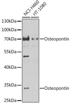
The antibody reacts with the 60 kDa osteopontin protein.

Immunogen

Epitope: aa 75-90
Synthetic peptide corresponding to aa 75-90 of osteopontin protein.

Application

Detect Osteopontin using this Anti-Osteopontin Antibody validated for use in WB, IH.
Research Category
Cell Structure
Research Sub Category
Cytoskeleton

Quality

Routinely evaluated by Western Blot on mouse kidney lysate.

Western Blot Analysis:
1:500 - 1:5,000 dilution of this antibody detected osteopontin in 10 μg of mouse kidney lysate.

Target description

60 kDa

Physical form

Serum containing 0.05% sodium azide.

Storage and Stability

Maintain at -20°C in undiluted aliquots for up to 1 year after date of receipt. Avoid repeated freeze/thaw cycles, which may damage IgG and affect product performance.

Analysis Note

Control
Mouse kidney, COS-1 and human urine lysate

Disclaimer

Unless otherwise stated in our catalog or other company documentation accompanying the product(s), our products are intended for research use only and are not to be used for any other purpose, which includes but is not limited to, unauthorized commercial uses, in vitro diagnostic uses, ex vivo or in vivo therapeutic uses or any type of consumption or application to humans or animals.

Not finding the right product?  

Try our Product Selector Tool.

Storage Class Code

10 - Combustible liquids

WGK

WGK 1


Certificates of Analysis (COA)

Search for Certificates of Analysis (COA) by entering the products Lot/Batch Number. Lot and Batch Numbers can be found on a product’s label following the words ‘Lot’ or ‘Batch’.

Already Own This Product?

Find documentation for the products that you have recently purchased in the Document Library.

Visit the Document Library

Kinga I Gawlik et al.
Scientific reports, 7, 44059-44059 (2017-03-11)
A large number of human diseases are caused by chronic tissue injury with fibrosis potentially leading to organ failure. There is a need for more effective anti-fibrotic therapies. Congenital muscular dystrophy type 1A (MDC1A) is a devastating form of muscular
Takahiro Fukaishi et al.
Diabetologia, 64(12), 2803-2816 (2021-09-10)
Pancreatic polypeptide (PP) cells, which secrete PP (encoded by the Ppy gene), are a minor population of pancreatic endocrine cells. Although it has been reported that the loss of beta cell identity might be associated with beta-to-PP cell-fate conversion, at
Silvia León et al.
Scientific reports, 6, 29073-29073 (2016-07-02)
Uterine growth and endometrial gland formation (adenogenesis) and function, are essential for fertility and are controlled by estrogens and other regulators, whose nature and physiological relevance are yet to be elucidated. Kisspeptin, which signals via Kiss1r, is essential for fertility
Fisetin lowers methylglyoxal dependent protein glycation and limits the complications of diabetes.
Maher, P; Dargusch, R; Ehren, JL; Okada, S; Sharma, K; Schubert, D
Testing null
Biomineralization in Three-Dimensional Scaffolds Based on Bacterial Nanocellulose for Bone Tissue Engineering: Feature Characterization and Stem Cell Differentiation.
Ca??as-Guti??rrez, et al.
Polymers, 15 (2023)

Our team of scientists has experience in all areas of research including Life Science, Material Science, Chemical Synthesis, Chromatography, Analytical and many others.

Contact Technical Service