Skip to Content
Merck
All Photos(9)

Key Documents

05-1000

Sigma-Aldrich

Anti-Phosphoserine Antibody, clone 4A4 (mouse IgG1)

clone 4A4, Upstate®, from mouse

Synonym(s):

Clone 4A4 Anti-Phosphoserine, Phosphoserine Detection Antibody

Sign Into View Organizational & Contract Pricing


About This Item

UNSPSC Code:
12352203
eCl@ss:
32160702
NACRES:
NA.41

biological source

mouse

Quality Level

antibody form

purified antibody

antibody product type

primary antibodies

clone

4A4, monoclonal

species reactivity

vertebrates

manufacturer/tradename

Upstate®

technique(s)

ELISA: suitable
flow cytometry: suitable
immunofluorescence: suitable
immunohistochemistry: suitable (paraffin)
western blot: suitable

isotype

IgG1

shipped in

dry ice

target post-translational modification

phosphorylation (pSer)

General description

The identification of protein phosphorylation as a regulatory mechanism originated from studies by Fischer and Krebs in the mid 1950s that later earned them the 1992 Nobel prize. It is the major mechanism for the regulation of diverse cellular processes including cell division, protein synthesis, transcriptional regulation and neurotransmission. The steady state phosphorylation of any given substrate is governed by the opposing activities of kinases and phosphatases. It is now believed that a third of all eukaryotic cellular proteins are phosphorylated and that the majority of all phosphorylation events occur on serine and threonine residues (>95%).

Specificity

This antibody recognizes serine-phosphorylated proteins from all species.

Immunogen

Phosphoserine coupled to KLH.

Application

Detect Phosphoserine using this Anti-Phosphoserine Antibody, clone 4A4 (mouse IgG1) validated for use in ELISA, FC, IF, IH(P) & WB.
Immunofluorescence

Flow Cytometry

Immunohistochemistry (Paraffin)

ELISA
Research Category
Signaling
Research Sub Category
General Post-translation Modification

Quality

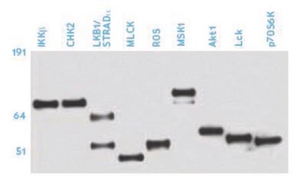
Routinely evaluated by Western Blot analysis on lysate from Calyculin A/Okadaic-treated human A431 carcinoma cells.

Western Blot Analysis:
0.5–2 μg/mL of this lot detected serinephosphorylated proteins in a lysate from either insulin or Calyculin A/Okadaic-treated human A431 carcinoma cells.

Target description

Dependent upon the molecular weight of the serine phosphorylated protein being detected.

Physical form

Format: Purified
Protein G-Sepharose Chromatography
Purified mouse monoclonal IgG1 in buffer containing PBS with 0.1% sodium azide and 30% glycerol.

Storage and Stability

Stable for 1 year at -20ºC from date of receipt.
For maximum recovery, centrifuge the original vial prior to cap removal. If the product has accidentally been frozen and thawed, spin it at 13,000 x g for 10 minutes at 2-8°C.

Other Notes

Concentration: Please refer to the Certificate of Analysis for the lot-specific concentration.

Legal Information

UPSTATE is a registered trademark of Merck KGaA, Darmstadt, Germany

Disclaimer

Unless otherwise stated in our catalog or other company documentation accompanying the product(s), our products are intended for research use only and are not to be used for any other purpose, which includes but is not limited to, unauthorized commercial uses, in vitro diagnostic uses, ex vivo or in vivo therapeutic uses or any type of consumption or application to humans or animals.

Not finding the right product?  

Try our Product Selector Tool.

Storage Class Code

12 - Non Combustible Liquids

WGK

WGK 2

Flash Point(F)

Not applicable

Flash Point(C)

Not applicable


Certificates of Analysis (COA)

Search for Certificates of Analysis (COA) by entering the products Lot/Batch Number. Lot and Batch Numbers can be found on a product’s label following the words ‘Lot’ or ‘Batch’.

Already Own This Product?

Find documentation for the products that you have recently purchased in the Document Library.

Visit the Document Library

Customers Also Viewed

Slide 1 of 1

1 of 1

Xiangfei Kong et al.
Journal of biochemistry, 146(3), 417-427 (2009-06-13)
CDK11(p58), a CDK11 family Ser/Thr kinase, is a G2/M specific protein and contributed to regulation of cell cycle, transcription and apoptotic signal transduction. Recently, CDK11(p58) has been reported to exert important functions in mitotic process, such as the regulation of
Increased NADPH oxidase-derived superoxide is involved in the neuronal cell death induced by hypoxia-ischemia in neonatal hippocampal slice cultures.
Lu, Q; Wainwright, MS; Harris, VA; Aggarwal, S; Hou, Y; Rau, T; Poulsen, DJ; Black, SM
Free radical biology & medicine null
Nucleocytoplasmic shuttling of a GATA transcription factor functions as a development timer.
Cai, H; Katoh-Kurasawa, M; Muramoto, T; Santhanam, B; Long, Y; Li, L; Ueda, M; Iglesias et al.
Science (New York, N.Y.) null
Glycine receptor internalization by protein kinases activation.
Miguel Angel Velazquez-Flores,Rocio Salceda,Miguel ??ngel Vel?!zquez-Flores,Roc?-o Salceda
Synapse null
Daisuke Nanba et al.
Stem cell research & therapy, 4(5), 127-127 (2014-01-11)
The development of an appropriate procedure for lentiviral gene transduction into keratinocyte stem cells is crucial for stem cell biology and regenerative medicine for genetic disorders of the skin. However, there is little information available on the efficiency of lentiviral

Our team of scientists has experience in all areas of research including Life Science, Material Science, Chemical Synthesis, Chromatography, Analytical and many others.

Contact Technical Service