Skip to Content
Merck
All Photos(5)

Documents

H9908

Sigma-Aldrich

Monoclonal Anti-phospho-Histone H3 (pSer28) antibody produced in rat

~0.5 mg/mL, clone HTA28, purified immunoglobulin, buffered aqueous solution

Synonym(s):

Monoclonal Anti-H3S28p

Sign Into View Organizational & Contract Pricing


About This Item

MDL number:
UNSPSC Code:
12352203
NACRES:
NA.41

biological source

rat

Quality Level

conjugate

unconjugated

antibody form

purified immunoglobulin

antibody product type

primary antibodies

clone

HTA28, monoclonal

form

buffered aqueous solution

mol wt

antigen 15 kDa

species reactivity

bovine, mouse, hamster, human

packaging

antibody small pack of 25 μL

concentration

~0.5 mg/mL

technique(s)

flow cytometry: suitable
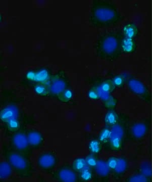
immunocytochemistry: suitable using 3.7% formaldehyde-methanol fixation
microarray: suitable
western blot: 0.5-1 μg/mL using whole extract of cultured human acute T cell leukemia Jurkat cells treated with Nocodazole.

isotype

IgG2a

UniProt accession no.

shipped in

dry ice

storage temp.

−20°C

target post-translational modification

phosphorylation (pSer28)

General description

Histone H1 and the N-terminal tail of histone-3 (H3) have key roles in the folding and inter-association of the chromatin fibers. Two currently known phosphorylation sites are present in the N-terminus of H3; serine-10 and serine-28. Mammals contain three main classes of histone H3 variants: the replicative histones (H3.1 and H3.2), the replacement histone (H3.3) and the centromeric histone (Cenp-A). Histone H3 is located on human chromosome 6p22.

Specificity

Monoclonal Anti-Phospho-Histone H3 (pSer28) reacts specifically with histone H3 phosphorylated at Ser28 near its N-terminus, and does not detect the unphosphorylated epitope. It detects the phosphorylated histone molecule at the onset of mitosis (prophase, metaphase and weaker at the beginning of anaphase), but not during late anaphase. Reactivity has been observed with human, bovine, hamster and mouse.
The antibody does not detect the unphosphorylated epitope. It detects the histone molecule at the onset of mitosis (prophase, metaphase, weaker at the beginning of anaphase, but not during late anaphase).

Immunogen

Synthetic peptide corresponding to amino acids of human histone H3.

Application

Monoclonal Anti-phospho-Histone H3 (pSer28) antibody is suitable for immunoblotting (approx. 15 kDa), immunocytochemistry (3.7% formaldehyde-methanol fixation) and flow cytometry applications.

Biochem/physiol Actions

Mitogenic stimulation, oncogenic transformation, or induction of oncogenic ras expression are accompanied with increase in serine-10 phosphorylation of the H3 N-terminal domain. It has been shown that phosphorylated H3 is associated with c-fos and c-myc genes in stimulated cells. Phosphorylation of H3, at both serine-10 and -28, coincides with the induction of mitotic chromosome condensation.
H3 phosphorylation may contribute to proto-oncogene induction by modulating chromatin structure and releasing blocks in elongation. H3 dephosphorylation occurs quite rapidly after mitosis and serine-10/28 reo main unphosphorylated throughout the remainder of interphase. PP1 has been identified as the H3 phosphatase.

Physical form

Solution in 0.01 M phosphate buffered saline, pH 7.4, containing 1% BSA and 15 mM sodium azide.

Preparation Note

Purified from culture supernatant of hybridoma cells grown in a bioreactor

Storage and Stability

For continuous use, store at 2 to 8 °C for up to one month. For extended storage, freeze in working aliquots. Repeated freezing and thawing is not recommended. Storage in "frost-free" freezers is not recommended. If slight turbidity occurs upon prolonged storage, clarify the solution by centrifugation before use. Working dilution samples should be discarded if not used within 12 hours.

Disclaimer

Unless otherwise stated in our catalog or other company documentation accompanying the product(s), our products are intended for research use only and are not to be used for any other purpose, which includes but is not limited to, unauthorized commercial uses, in vitro diagnostic uses, ex vivo or in vivo therapeutic uses or any type of consumption or application to humans or animals.

Not finding the right product?  

Try our Product Selector Tool.

Storage Class Code

10 - Combustible liquids

WGK

WGK 3

Flash Point(F)

Not applicable

Flash Point(C)

Not applicable


Certificates of Analysis (COA)

Search for Certificates of Analysis (COA) by entering the products Lot/Batch Number. Lot and Batch Numbers can be found on a product’s label following the words ‘Lot’ or ‘Batch’.

Already Own This Product?

Find documentation for the products that you have recently purchased in the Document Library.

Visit the Document Library

An FGF-WNT gene regulatory network controls lung mesenchyme development.
Yin Y, et al.
Developmental Biology, 319(2) (2008)
Molecular basis for the recognition of phosphorylated and phosphoacetylated histone h3 by 14-3-3.
Macdonald N, et al.
Molecular Cell, 20(2) (2005)
CREB controls MSK1-mediated phosphorylation of histone H3 at the c-fos promoter in vitro.
Shimada M, et al.
The Journal of Biological Chemistry, M109-M109 (2010)
Majd Haj et al.
The Journal of biological chemistry, 292(50), 20720-20731 (2017-10-22)
Pituitary gonadotropin hormones are regulated by gonadotropin-releasing hormone (GnRH) via MAPK signaling pathways that stimulate gene transcription of the common α-subunit (Cga) and the hormone-specific β-subunits of gonadotropin. We have reported previously that GnRH-induced activities at these genes include various
Mutations in QARS, encoding glutaminyl-tRNA synthetase, cause progressive microcephaly, cerebral-cerebellar atrophy, and intractable seizures.
Zhang X, et al.
American Journal of Human Genetics, 94(4) (2014)

Our team of scientists has experience in all areas of research including Life Science, Material Science, Chemical Synthesis, Chromatography, Analytical and many others.

Contact Technical Service