Skip to Content
Merck
All Photos(1)

Documents

428017

Sigma-Aldrich

Anti-Lamp1 Mouse mAb (LY1C6)

liquid, clone LY1C6, Calbiochem®

Synonym(s):

Anti-Lysosome-Associated Membrane Protein 1

Sign Into View Organizational & Contract Pricing


About This Item

UNSPSC Code:
12352203
NACRES:
NA.41

biological source

mouse

Quality Level

antibody form

purified antibody

antibody product type

primary antibodies

clone

LY1C6, monoclonal

form

liquid

contains

≤0.09% sodium azide as preservative

species reactivity

rat

manufacturer/tradename

Calbiochem®

storage condition

OK to freeze
avoid repeated freeze/thaw cycles

isotype

IgG1

shipped in

wet ice

storage temp.

−20°C

target post-translational modification

unmodified

Gene Information

General description

Protein G purified mouse monoclonal antibody. Recognizes the ~120 kDa Lamp1 protein.
Recognizes the ~120 kDa Lamp1 protein.
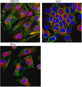
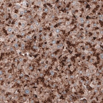
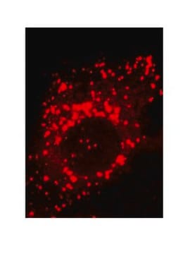
This Anti-Lamp1 Mouse mAb (LY1C6) is validated for use in Immunoblotting, Immunocytochemistry, Immunofluorescence, Immunoprecipitation for the detection of Lamp1.

Immunogen

Rat
rat liver lysosomal membranes

Application

Immunoblotting (1 µg/ml, chemiluminescence)

Immunocytochemistry (1:100)

Immunofluorescence (see comments)

Immunoprecipitation (see comments)

Packaging

Please refer to vial label for lot-specific concentration.

Warning

Toxicity: Standard Handling (A)

Physical form

In PBS containing 50% glycerol, pH 7.2.

Reconstitution

Following initial thaw, aliquot and freeze (-20°C).

Analysis Note

Positive Control
CHO-K1 cells

Other Notes

Kannan, K. et al. 1996. Cell Immunol.171, 10.
Rohrer, J., et al. 1996. J. Cell Biol. 132, 565.
Howe et al. 1988. PNAS85, 7577.
Lewis et al. 1985. J. Cell Biol.100, 1839.
This antibody has also been reported to work for immunofluorescence and immunoprecipitation. Antibody should be titrated for optimal results in individual systems.

Legal Information

CALBIOCHEM is a registered trademark of Merck KGaA, Darmstadt, Germany

Not finding the right product?  

Try our Product Selector Tool.

Storage Class Code

10 - Combustible liquids

WGK

WGK 1

Flash Point(F)

Not applicable

Flash Point(C)

Not applicable


Certificates of Analysis (COA)

Search for Certificates of Analysis (COA) by entering the products Lot/Batch Number. Lot and Batch Numbers can be found on a product’s label following the words ‘Lot’ or ‘Batch’.

Already Own This Product?

Find documentation for the products that you have recently purchased in the Document Library.

Visit the Document Library

Arvind Chhabra et al.
European journal of immunology, 34(10), 2824-2833 (2004-09-16)
Dendritic cells (DC) capture antigens from apoptotic and/or necrotic tumor cells and cross-present them to T cells, and various ways of delivering tumor antigens to DC in vitro and in vivo are being pursued. Since fusions of antigenic proteins with
Vanessa Ginet et al.
The American journal of pathology, 175(5), 1962-1974 (2009-10-10)
The multiplicity of cell death mechanisms induced by neonatal hypoxia-ischemia makes neuroprotective treatment against neonatal asphyxia more difficult to achieve. Whereas the roles of apoptosis and necrosis in such conditions have been studied intensively, the implication of autophagic cell death
María Morell et al.
EBioMedicine, 76, 103808-103808 (2022-01-23)
Type I IFN (IFN-I) is a family of cytokines involved in the pathogenesis of autoimmune and autoinflammatory diseases such as psoriasis. SIDT1 is an ER-resident protein expressed in the lymphoid lineage, and involved in anti-viral IFN-I responses in vivo, through
Raymond E Hulse et al.
The Journal of neuroscience : the official journal of the Society for Neuroscience, 28(47), 12199-12211 (2008-11-21)
In brain, monomeric immunoglobin G (IgG) is regarded as quiescent and only poised to initiate potentially injurious inflammatory reactions via immune complex formation associated with phagocytosis and tumor necrosis factor alpha (TNF-alpha) production in response to disease. Using rat hippocampal
Julien Puyal et al.
Annals of neurology, 66(3), 378-389 (2009-06-25)
To evaluate the contributions of autophagic, necrotic, and apoptotic cell death mechanisms after neonatal cerebral ischemia and hence define the most appropriate neuroprotective approach for postischemic therapy. Rats were exposed to transient focal cerebral ischemia on postnatal day 12. Some

Articles

Autophagy is a regulated process involved in cell growth, development, and recycling of cytoplasmic components in cells.

Autophagy is a regulated process involved in cell growth, development, and recycling of cytoplasmic components in cells.

Autophagy is a regulated process involved in cell growth, development, and recycling of cytoplasmic components in cells.

Autophagy is a regulated process involved in cell growth, development, and recycling of cytoplasmic components in cells.

Our team of scientists has experience in all areas of research including Life Science, Material Science, Chemical Synthesis, Chromatography, Analytical and many others.

Contact Technical Service