Skip to Content
Merck
All Photos(1)

Documents

248M-9

Sigma-Aldrich

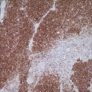
Ep-CAM/Epithelial Specific Antigen (Ber-EP4) Mouse Monoclonal Antibody

Sign Into View Organizational & Contract Pricing


About This Item

UNSPSC Code:
12352203
NACRES:
NA.41

biological source

mouse

Quality Level

100
500

conjugate

unconjugated

antibody form

culture supernatant

antibody product type

primary antibodies

clone

Ber-EP4, monoclonal

description

For In Vitro Diagnostic Use in Select Regions (See Chart)

form

buffered aqueous solution

species reactivity

human

packaging

vial of 0.1 mL concentrate (248M-94)
vial of 0.5 mL concentrate (248M-95)
bottle of 1.0 mL predilute (248M-97)
vial of 1.0 mL concentrate (248M-96)
bottle of 7.0 mL predilute (248M-98)

manufacturer/tradename

Cell Marque

technique(s)

immunohistochemistry (formalin-fixed, paraffin-embedded sections): 1:50-1:200

isotype

IgG1κ

control

colon carcinoma

shipped in

wet ice

storage temp.

2-8°C

visualization

cytoplasmic

Gene Information

human ... EPCAM(4072)

General description

Epithelial cell adhesion molecule (Ep-CAM) is a transmembrane glycoprotein localized on the membrane of cells in most epithelial tissues. Immunoreactivity with the antibody to E-CAM has been seen in the majority of epithelial neoplasms, whereas most non-epithelial neoplasms do not show Ep-CAM expression. Ep-CAM is not expressed in mesothelial cells, hepatocytes, and lymphocytes. In conjunction with other markers, Ep-CAM can be used as an aid in determining the epithelial origin of neoplasms such as lung adenocarcinoma.

Quality


IVD

IVD

IVD

RUO

Linkage

Ep-CAM Positive Control Slides, Product No. 248S, are available for immunohistochemistry (formalin-fixed, paraffin-embedded sections).

Physical form

Solution in Tris Buffer, pH 7.3-7.7, with 1% BSA and <0.1% Sodium Azide

Preparation Note

Download the IFU specific to your product lot and formatNote: This requires a keycode which can be found on your packaging or product label.

Other Notes

For Technical Service please contact: 800-665-7284 or email: service@cellmarque.com

Legal Information

Cell Marque is a trademark of Merck KGaA, Darmstadt, Germany

Not finding the right product?  

Try our Product Selector Tool.


Certificates of Analysis (COA)

Search for Certificates of Analysis (COA) by entering the products Lot/Batch Number. Lot and Batch Numbers can be found on a product’s label following the words ‘Lot’ or ‘Batch’.

Already Own This Product?

Find documentation for the products that you have recently purchased in the Document Library.

Visit the Document Library

Nelson G Ordóñez
Advances in anatomic pathology, 13(1), 16-25 (2006-02-08)
At present, a large number of immunohistochemical markers that can be used in the differential diagnosis between epithelioid peritoneal mesotheliomas and serous carcinomas are available. However, great differences of opinion exist regarding the individual value of some of these markers.
Nelson G Ordóñez
Modern pathology : an official journal of the United States and Canadian Academy of Pathology, Inc, 19(3), 417-428 (2006-01-18)
As both mesotheliomas and squamous carcinomas can present a wide variety of morphological patterns, they can on occasion be confused. Recently, some groups of investigators have called attention to the difficulties that sometimes exist in distinguishing between these malignancies and
U Latza et al.
Journal of clinical pathology, 43(3), 213-219 (1990-03-01)
A new monoclonal antibody, Ber-EP4, directed against a partially formol resistant epitope on the protein moiety of two 34 kilodalton and 39 kilodalton glycopolypeptides on human epithelial cells is described. Immunostaining of a wide range of normal and neoplastic human
N G Ordóñez
American journal of clinical pathology, 109(1), 85-89 (1998-01-14)
Although most studies have indicated that Ber-EP4 immunostaining can assist in differentiating epithelial pleural mesotheliomas from adenocarcinomas that metastasize to the pleura, the percentage of positive cases has varied greatly among different studies. Authors of a recent publication concluded that
C K Ma et al.
American journal of clinical pathology, 99(5), 551-557 (1993-05-01)
Distinguishing primary hepatocellular carcinoma (HCC) from metastatic carcinomas to the liver is often difficult, if not impossible, particularly in needle biopsy and fine-needle aspiration specimens. In an attempt to identify a specific immunohistochemical profile that would distinguish HCC from metastatic

Related Content

Diagnostic immunohistochemistry overview highlights its importance in cancer diagnosis and detecting infectious diseases in modern clinical pathology.

Diagnostic immunohistochemistry overview highlights its importance in cancer diagnosis and detecting infectious diseases in modern clinical pathology.

Diagnostic immunohistochemistry overview highlights its importance in cancer diagnosis and detecting infectious diseases in modern clinical pathology.

Diagnostic immunohistochemistry overview highlights its importance in cancer diagnosis and detecting infectious diseases in modern clinical pathology.

Our team of scientists has experience in all areas of research including Life Science, Material Science, Chemical Synthesis, Chromatography, Analytical and many others.

Contact Technical Service