Skip to Content
Merck
All Photos(1)

Key Documents

MAB367

Sigma-Aldrich

Anti-Muscarinic Acetylcholine Receptor m2 Antibody, clone M2-2-B3

clone M2-2-B3, Chemicon®, from rat

Synonym(s):

Muscarinic Receptor Antibody

Sign Into View Organizational & Contract Pricing


About This Item

UNSPSC Code:
12352203
eCl@ss:
32160702
NACRES:
NA.41

biological source

rat

Quality Level

antibody form

purified immunoglobulin

antibody product type

primary antibodies

clone

M2-2-B3, monoclonal

species reactivity

human, monkey, rat

species reactivity (predicted by homology)

mammals

manufacturer/tradename

Chemicon®

technique(s)

immunocytochemistry: suitable
immunohistochemistry: suitable
immunoprecipitation (IP): suitable

isotype

IgG2a

suitability

not suitable for immunohistochemistry (Paraffin)

UniProt accession no.

shipped in

wet ice

target post-translational modification

unmodified

Gene Information

human ... CHRM2(1129)

Specificity

m2 muscarinic acetylcholine receptor. No reactivity with the other subtypes.

SPECIES REACTIVITIES:

Expected to react with most mammalian species.

Immunogen

i3 loop of m2 receptor fusion protein (225-359), fused to Glutathione S-transferase.

Application

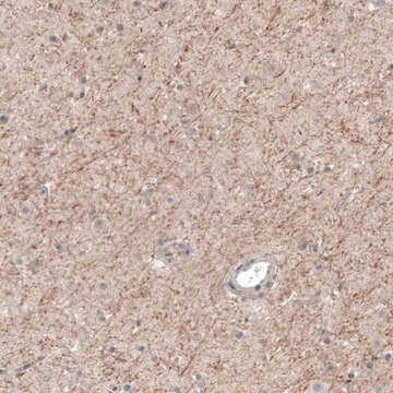
Anti-Muscarinic Acetylcholine Receptor m2 Antibody, clone M2-2-B3 is an antibody against Muscarinic Acetylcholine Receptor m2 for use in IC, IH & IP.
Immunohistochemistry on 4% paraformaldehyde fixed tissue. Does not work on paraffin embedded tissues. Suggested starting concentration 1-5 μg/mL. It is suggested that you use the PAP system if using this antibody on rat. Immunocytochemistry on transfected cells

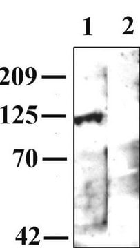
Immunoprecipitation Works poorly for immunoblotting Optimal working dilutions must be determined by end user.

IMMUNOHISTOCHEMISTRY PROTOCOL FOR MAB367

This antibody has been used successfully on 30 mm, free floating, 4% paraformaldehyde fixed rat brain tissue. All steps are performed under constant agitation. Suggested protocol follows.

1) 3 x 10 minute washes in TBS (with or without 0.25% Triton).

2) Incubate for 30 minutes in TBS with 3% serum (same as host from secondary antibody).

3) Incubate primary antibody diluted appropriately in TBS with 1% serum (same as host from secondary antibody) (with or without 0.25% Triton) for 2 hours at room temperature followed by 16 hours at 4°C.

4) 3 x 10 minute washes in TBS.

5) Incubate with secondary antibody diluted appropriately in TBS with 1% serum (same as host from secondary antibody).

6) 3 x 10 minute washes in TBS.

7) ABC Elite (1:200 Vector Labs) in TBS.

8) 2 x 10 minute washes in TBS.

9) 1 x 10 minute wash in phosphate buffer (no saline).

10) DAB reaction with 0.06% NiCl added for intensification.

11) 2 x 10 minute washes in PBS.

12) 1 x 10 minute wash in phosphate buffer (no saline).
Research Category
Neuroscience
Research Sub Category
Neurotransmitters & Receptors

Physical form

Format: Purified
Purified immunoglobulin. Liquid in 0.02 M phosphate buffer, 0.25 M NaCl with 0.1% sodium azide, pH 7.6.

Storage and Stability

Maintain at 2-8°C in undiluted aliquots for up to 6 months.

Other Notes

Concentration: Please refer to the Certificate of Analysis for the lot-specific concentration.

Legal Information

CHEMICON is a registered trademark of Merck KGaA, Darmstadt, Germany

Disclaimer

Unless otherwise stated in our catalog or other company documentation accompanying the product(s), our products are intended for research use only and are not to be used for any other purpose, which includes but is not limited to, unauthorized commercial uses, in vitro diagnostic uses, ex vivo or in vivo therapeutic uses or any type of consumption or application to humans or animals.

Not finding the right product?  

Try our Product Selector Tool.

Storage Class Code

10 - Combustible liquids

WGK

WGK 2

Flash Point(F)

Not applicable

Flash Point(C)

Not applicable


Certificates of Analysis (COA)

Search for Certificates of Analysis (COA) by entering the products Lot/Batch Number. Lot and Batch Numbers can be found on a product’s label following the words ‘Lot’ or ‘Batch’.

Already Own This Product?

Find documentation for the products that you have recently purchased in the Document Library.

Visit the Document Library

Lisa Lambert et al.
Frontiers in cellular neuroscience, 12, 450-450 (2018-12-18)
Our aim was to examine the dynamics of the muscarinic m2 receptor (m2R), a G-protein coupled receptor (GPCR), after agonist activation in living hippocampal neurons, and especially clathrin dependency endocytosis. We have previously shown that the m2R undergoes agonist-induced internalization
Roles of M2 and M4 muscarinic receptors in regulating acetylcholine release from myenteric neurons of mouse ileum.
Takeuchi, T; Fujinami, K; Goto, H; Fujita, A; Taketo, MM; Manabe, T; Matsui, M; Hata, F
Journal of Neurophysiology null
Maria Medalla et al.
The Journal of neuroscience : the official journal of the Society for Neuroscience, 32(44), 15611-15625 (2012-11-02)
The anterior cingulate cortex (ACC) and dorsolateral prefrontal cortices (DLPFC) share robust excitatory connections. However, during rapid eye movement (REM) sleep, when cortical activity is dominated by acetylcholine, the ACC is activated but DLPFC is suppressed. Using pathway tracing and
Rinaldo David D'Souza et al.
eLife, 5 (2016-10-22)
Diverse features of sensory stimuli are selectively processed in distinct brain areas. The relative recruitment of inhibitory and excitatory neurons within an area controls the gain of neurons for appropriate stimulus coding. We examined how such a balance of inhibition
Răzvan Gămănuţ et al.
Neuron, 97(3), 698-715 (2018-02-09)
The inter-areal wiring pattern of the mouse cerebral cortex was analyzed in relation to a refined parcellation of cortical areas. Twenty-seven retrograde tracer injections were made in 19 areas of a 47-area parcellation of the mouse neocortex. Flat mounts of

Our team of scientists has experience in all areas of research including Life Science, Material Science, Chemical Synthesis, Chromatography, Analytical and many others.

Contact Technical Service