Skip to Content
Merck
All Photos(2)

Documents

C7930

Sigma-Aldrich

Anti-CD3, T Cell antibody produced in rabbit

whole antiserum

Synonym(s):

Anti-CD3epsilon, Anti-IMD18, Anti-T3E, Anti-TCRE

Sign Into View Organizational & Contract Pricing


About This Item

MDL number:
UNSPSC Code:
12352203
NACRES:
NA.41

biological source

rabbit

conjugate

unconjugated

antibody form

whole antiserum

antibody product type

primary antibodies

clone

polyclonal

mol wt

antigen 20 kDa (bovine and swine)

contains

15 mM sodium azide

species reactivity

rat, canine, mouse, human, Psammomys (sand rat), sheep, feline, pig, chicken, bovine

technique(s)

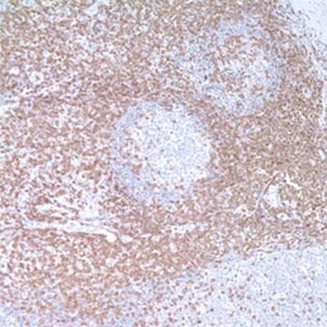
immunohistochemistry (formalin-fixed, paraffin-embedded sections): 1:200 using enzyme pre-digested, formalin-fixed, paraffin-embedded human tonsil sections
western blot: 1:200 using mouse thymus extract

UniProt accession no.

shipped in

dry ice

storage temp.

−20°C

target post-translational modification

unmodified

Gene Information

human ... CD3E(916)
mouse ... Cd3e(12501)
rat ... Cd3e(315609)

General description

CD3 is a 20kDa glycoprotein expressed on surface of all human T lymphocytes. CD3 molecule is encoded by the gene mapped to human chromosome 11q23.3. The CD3 antigen is expressed during thymopoiesis. It is also present in the majority of T-CLL and T-lymphomas and in approximately 70% of T-ALL.

Specificity

The antibody reacts with the proline-rich peptide sequence comprising 13 amino acids in the cytoplasmic domain of the CD3-ε chain. It detects normal, reactive and most neoplastic human T cells in routinely processed enzyme pre-digested, formalin-fixed, paraffin-embedded tissue sections. The antibody also reacts with human tissues fixed with Bouin′s solution and embedded in paraffin. Frozen sections and smears are also suitable for staining with this antibody. The antibody reacts with T cell areas in lymphoid tissues derived from various species.

Immunogen

synthetic human CD3 peptide (residues 156-168 of the CD3-ε chain) coupled to porcine thyroglobulin. Coupling was performed by a modification of the reported method.

Application

Anti-CD3, T Cell antibody produced in rabbit has been used in:
  • immunoblotting
  • immunofluorescence staining
  • immunohistochemistry
  • immunohistochemical analysis

Biochem/physiol Actions

CD3 is a human cell surface glycoprotein complex involved in signal transduction following binding of antigen receptor.

Disclaimer

Unless otherwise stated in our catalog or other company documentation accompanying the product(s), our products are intended for research use only and are not to be used for any other purpose, which includes but is not limited to, unauthorized commercial uses, in vitro diagnostic uses, ex vivo or in vivo therapeutic uses or any type of consumption or application to humans or animals.

Not finding the right product?  

Try our Product Selector Tool.

Storage Class Code

12 - Non Combustible Liquids

WGK

nwg

Flash Point(F)

Not applicable

Flash Point(C)

Not applicable


Certificates of Analysis (COA)

Search for Certificates of Analysis (COA) by entering the products Lot/Batch Number. Lot and Batch Numbers can be found on a product’s label following the words ‘Lot’ or ‘Batch’.

Already Own This Product?

Find documentation for the products that you have recently purchased in the Document Library.

Visit the Document Library

Spontaneous Mammary Tumors Differ Widely in Their Inherent Sensitivity to Adoptively Transferred T Cells
Wall EM, et al.
Cancer research, 67(13), 6442-6450 (2007)
The T cell receptor/CD3 complex: a dynamic protein ensemble
Clevers H, et al.
Annual Review of Immunology, 6(1), 629-662 (1988)
Nadir Kadri et al.
Journal of immunology (Baltimore, Md. : 1950), 188(7), 3138-3149 (2012-03-01)
Type 1 diabetes (T1D) is a chronic autoimmune disease that results from T cell-mediated destruction of pancreatic β cells. CD1d-restricted NKT lymphocytes have the ability to regulate immunity, including autoimmunity. We previously demonstrated that CD1d-restricted type II NKT cells, which
Chuanhai Cao et al.
Neurobiology of disease, 34(1), 63-70 (2009-01-27)
We have found that a small number of purified Th2-biased Abeta-specific T cells are sufficient to provide profound cognitive and pathological benefits in an APP+PS1 mouse model for Alzheimer's disease. Six weeks after receiving T cell infusions, cognitively-impaired mice performed
Fabian P S Yu et al.
Scientific reports, 8(1), 1808-1808 (2018-01-31)
Farber Disease (FD) is an ultra-rare Lysosomal Storage Disorder caused by deficient acid ceramidase (ACDase) activity. Patients with ACDase deficiency manifest a spectrum of symptoms including formation of nodules, painful joints, and a hoarse voice. Classic FD patients will develop

Our team of scientists has experience in all areas of research including Life Science, Material Science, Chemical Synthesis, Chromatography, Analytical and many others.

Contact Technical Service