Skip to Content
Merck
All Photos(1)

Documents

04-135

Sigma-Aldrich

Anti-Brachyury Antibody, clone 3E4.2

clone 3E4.2, Upstate®, from mouse

Synonym(s):

T brachyury (mouse) homolog, T brachyury-like, T protein, T, brachyury homolog (mouse), transcription factor T

Sign Into View Organizational & Contract Pricing


About This Item

UNSPSC Code:
12352203
eCl@ss:
32160702
NACRES:
NA.41

biological source

mouse

Quality Level

antibody form

purified immunoglobulin

antibody product type

primary antibodies

clone

3E4.2, monoclonal

species reactivity

human

manufacturer/tradename

Upstate®

technique(s)

western blot: suitable

input

sample type mesenchymal stem cell(s)

isotype

IgG1κ

NCBI accession no.

UniProt accession no.

shipped in

wet ice

target post-translational modification

unmodified

Gene Information

human ... TBXT(6862)

General description

The Brachyury protein is a T-box transcription factor that is one of the earliest indicators of mesoderm formation during embryonic development. As the brachyury protein family is essential for the formation of the posterior body in all vertebrates, Brachyury has been shown to be expressed during gastrula and neurula stages, as well as in the developing notochord. Consequently, when mutations occur within the brachyury gene, severe vertebral malformations have been found to occur, as a direct result. In addition, current findings also indicate that the dysregulation of brachyury may also be linked to the formation of chordomas, which are malignant tumors that develop along the spine.

Specificity

Reactivity with other species has not been tested.
This monoclonal antibody recognizes the brachyury protein.

Immunogen

Epitope: C-terminus
Synthetic human brachyury peptide, a.a. 224-435.

Application

Research Category
Stem Cell Research
Research Sub Category
Mesenchymal Stem Cells
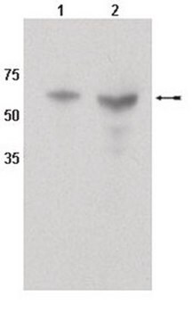
This Anti-Brachyury Antibody, clone 3E4.2 is validated for use in WB for the detection of Brachyury.

Quality

Routinely evaluated by Western blot on NT2 cell lysates.

Western Blotting:
Recommended working dilutions are 1:1000.

Target description

47 kDa

Physical form

Format: Purified
Protein G Purified
Purified mouse monoclonal IgG1κ in buffer containing PBS with 0.05% sodium azide.

Storage and Stability

Maintain refrigerated at 2-8°C in undiluted aliquots for up to 12 months. Avoid repeat freeze/thaw cycles.

Other Notes

Concentration: Please refer to the Certificate of Analysis for the lot-specific concentration.

Legal Information

UPSTATE is a registered trademark of Merck KGaA, Darmstadt, Germany

Disclaimer

Unless otherwise stated in our catalog or other company documentation accompanying the product(s), our products are intended for research use only and are not to be used for any other purpose, which includes but is not limited to, unauthorized commercial uses, in vitro diagnostic uses, ex vivo or in vivo therapeutic uses or any type of consumption or application to humans or animals.

Not finding the right product?  

Try our Product Selector Tool.

Storage Class Code

12 - Non Combustible Liquids

WGK

WGK 1

Flash Point(F)

Not applicable

Flash Point(C)

Not applicable


Certificates of Analysis (COA)

Search for Certificates of Analysis (COA) by entering the products Lot/Batch Number. Lot and Batch Numbers can be found on a product’s label following the words ‘Lot’ or ‘Batch’.

Already Own This Product?

Find documentation for the products that you have recently purchased in the Document Library.

Visit the Document Library

T P Yamaguchi et al.
Genes & development, 13(24), 3185-3190 (2000-01-05)
Wnt3a encodes a signal that is expressed in the primitive streak of the gastrulating mouse embryo and is required for paraxial mesoderm development. In its absence cells adopt ectopic neural fates. Embryos lacking the T-box-containing transcription factors, Brachyury or Tbx6
Brachyury, a crucial regulator of notochordal development, is a novel biomarker for chordomas.
Vujovic, S, et al.
The Journal of Pathology, 209, 157-165 (2006)
Development of antibodies to human embryonic stem cell antigens.
Cai, Jingli, et al.
BMC Developmental Biology, 5, 26-26 (2005)
Hematopoiesis from human embryonic stem cells: overcoming the immune barrier in stem cell therapies.
Priddle, Helen, et al.
Stem Cells, 24, 815-824 (2006)
Chromatin remodeling during mouse and human embryonic stem cell differentiation.
Golob, Jonathan L, et al.
Developmental Dynamics, 237, 1389-1398 (2008)

Our team of scientists has experience in all areas of research including Life Science, Material Science, Chemical Synthesis, Chromatography, Analytical and many others.

Contact Technical Service