General description
Histone H3.1 (UniProt: P68431; also known as Histone H3/a, Histone H3/b, Histone H3/c, Histone H3/d, Histone H3/f, Histone H3/h, Histone H3/I, Histone H3/j, Histone H3/k, Histone H3/l) is encoded by the HIST1H3A (also known as H3FA, HIST1H3B, H3FL, HIST1H3C, H3FC, HIST1H3D, H3FB, HISTH3F, H3FD, HIST1H3F, H3FI, HIST1H3G, H3FH, HIST1H3H, H3FK, HIST1H3I, H3FF, HIST1H3J, H3FJ) gene (Gene ID: 8350, 8351, 8352, 8354, 8355, 8356, 8357, 8358, 8968) in human. Histone H3 has two main variants, H3.1 and H3.3, which show different genomic localization patterns in animals. The H3.1 and H3.3 complexes also possess distinct histone chaperones, CAF-1 and HIRA, which play important role in mediating DNA-synthesis-dependent and -independent nucleosome assembly. It has been reported that Histone H3.1 serves as the canonical histone, which is incorporated during DNA replication, whereas H3.3 acts as the replacement histone that can be incorporated outside of S-phase during chromatin-disrupting processes like transcription. Histone H 3.1 is a core component of nucleosome that is present only in mammals and is usually enriched in acetylation of Lysine 15 and demethylation of lysine 10 (HeK9Me2). It is expressed during S phase, then expression decreases significantly as cell division slows down during the process of differentiation. Histone H 3.1 expression is shown to be replication dependent. It s presence at the site of UV-induced DNA damage has also been reported. It has also been shown that H3.1/H4 tetramers do not split and remain intact during replication dependent deposition of H3.1 variant. (Ref.: Stroud, H., et al (2012). Proc. Natl. Acad. Sci. USA 109(14); 5370-5375).
Specificity
This rabbit polyclonal antibody detects Histone H3 in human and murine cells.
Immunogen
Epitope: C-terminus
KLH-conjugated linear peptide corresponding to 12 amino acids from the C-terminus of human Histone H3.1.
Application
Anti-Histone H3, CT, pan, Cat. No. ABE865, is a highly specific rabbit polyclonal antibody that targets Histone H3.1 and has been tested in Immunocytochemistry and Western Blotting.
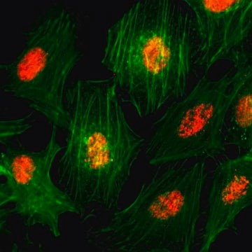
Immunocytochemistry Analysis: A 1:100 dilution from a representative lot detected Histone H3, CT, pan in HeLa and NIH/3T3 cells.
Research Category
Epigenetics & Nuclear Function
Quality
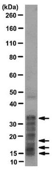
Evaluated by Western Blotting in HeLa cell acid extract.
Western Blotting Analysis: A 1:5,000 dilution of this antibody detected Histone H3, CT, pan in HeLa cell acid extract.
Target description
~17 kDa observed; 15.40 kDa calculated. Uncharacterized bands may be observed in some lysate(s).
Physical form
Affinity Purified
Purified rabbit polyclonal antibody in buffer containing 0.1 M Tris-Glycine (pH 7.4), 150 mM NaCl with 0.05% sodium azide.
Storage and Stability
Stable for 1 year at 2-8°C from date of receipt.
Other Notes
Concentration: Please refer to lot specific datasheet.
Disclaimer
Unless otherwise stated in our catalog or other company documentation accompanying the product(s), our products are intended for research use only and are not to be used for any other purpose, which includes but is not limited to, unauthorized commercial uses, in vitro diagnostic uses, ex vivo or in vivo therapeutic uses or any type of consumption or application to humans or animals.