Skip to Content
Merck
All Photos(3)

Documents

ABC99

Sigma-Aldrich

Anti-SFTPC Antibody

from rabbit, purified by affinity chromatography

Synonym(s):

Pulmonary surfactant-associated protein C, SP-C, Pulmonary surfactant-associated proteolipid SPL(Val), SP5

Sign Into View Organizational & Contract Pricing


About This Item

UNSPSC Code:
12352203
eCl@ss:
32160702
NACRES:
NA.41

biological source

rabbit

Quality Level

antibody form

affinity isolated antibody

antibody product type

primary antibodies

clone

polyclonal

purified by

affinity chromatography

species reactivity

mouse, rat, human

technique(s)

immunohistochemistry: suitable (paraffin)
western blot: suitable

NCBI accession no.

UniProt accession no.

shipped in

wet ice

target post-translational modification

unmodified

Gene Information

human ... SFTPC(6440)

General description

SFTPC, also known as SFTP2 and Pulmonary surfactant-associated protein C, is a complex mixture of phospholipids and proteins secreted from airway epithelial cells (type II cells) in alveoli which reduces the surface tension at the alveolar air liquid interface, providing alveolar stability necessary for normal ventilation. Four distinct proteins isolated from pulmonary surfactant are termed surfactant proteins A, B, C, and D. SP A (28 - 36 kDa) and SP D (43 kDa) are collagenous carbohydrate binding proteins, whereas SP B (8 - 9 kDa) and SP C (4 kDa) are non collagenous hydrophobic proteins. SP C is a fragment of a much larger precursor protein of 21 kDa. The precursor contains an exceedingly hydrophobic region of 34 amino acids that comprises most of the mature SP C, and a unique poly-valine domain.

Immunogen

Epitope: N-terminus
KLH-conjugated linear peptide corresponding to the N-terminus of human SFTPC.

Application

Detect SP-C using this rabbit polyclonal antibody, Anti-SFTPC Antibody validated for use in western blotting & IHC (Paraffin).
Immunohistochemistry Analysis: A 1:500 dilution from a representative lot detected SFTPC in human lung tissue.
Research Category
Apoptosis & Cancer
Research Sub Category
Apoptosis - Additional

Quality

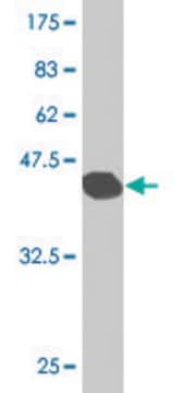
Evaluated by Western Blotting in rat lung tissue lysate.

Western Blotting Analysis: 1.0 µg/mL of this antibody detected SFTPC in 10 µg of rat lung tissue lysate.

Target description

~20 kDa observed

Physical form

Affinity purified
Purified rabbit polyclonal in buffer containing 0.1 M Tris-Glycine (pH 7.4), 150 mM NaCl with 0.05% sodium azide.

Storage and Stability

Stable for 1 year at 2-8°C from date of receipt.

Analysis Note

Control
Rat lung tissue lysate

Other Notes

Concentration: Please refer to the Certificate of Analysis for the lot-specific concentration.

Disclaimer

Unless otherwise stated in our catalog or other company documentation accompanying the product(s), our products are intended for research use only and are not to be used for any other purpose, which includes but is not limited to, unauthorized commercial uses, in vitro diagnostic uses, ex vivo or in vivo therapeutic uses or any type of consumption or application to humans or animals.

Not finding the right product?  

Try our Product Selector Tool.

Storage Class Code

12 - Non Combustible Liquids

WGK

WGK 1

Flash Point(F)

Not applicable

Flash Point(C)

Not applicable


Certificates of Analysis (COA)

Search for Certificates of Analysis (COA) by entering the products Lot/Batch Number. Lot and Batch Numbers can be found on a product’s label following the words ‘Lot’ or ‘Batch’.

Already Own This Product?

Find documentation for the products that you have recently purchased in the Document Library.

Visit the Document Library

William J Zacharias et al.
Nature, 555(7695), 251-255 (2018-03-01)
Functional tissue regeneration is required for the restoration of normal organ homeostasis after severe injury. Some organs, such as the intestine, harbour active stem cells throughout homeostasis and regeneration; more quiescent organs, such as the lung, often contain facultative progenitor
Shanru Li et al.
Stem cell reports, 18(9), 1841-1853 (2023-08-19)
AT2 cells harbor alveolar stem cell activity in the lung and can self-renew and differentiate into AT1 cells during homeostasis and after injury. To identify epigenetic pathways that control the AT2-AT1 regenerative response in the lung, we performed an organoid
Santiago Naranjo et al.
Genes & development, 36(15-16), 936-949 (2022-09-30)
Lung cancer is the leading cause of cancer-related death worldwide. Lung adenocarcinoma (LUAD), the most common histological subtype, accounts for 40% of all cases. While existing genetically engineered mouse models (GEMMs) recapitulate the histological progression and transcriptional evolution of human
Mariyan J Jeyarajah et al.
Proceedings of the National Academy of Sciences of the United States of America, 119(49), e2203071119-e2203071119 (2022-11-29)
Remodeling of the uterine vasculature by invasive extravillous trophoblasts (EVTs) is a critical aspect of human placentation. Insufficient EVT invasion can lead to severe obstetrical complications like preeclampsia, intrauterine growth restriction, and preterm birth. Glial cells missing-1 (GCM1) is a
Kuei-Pin Chung et al.
Nature communications, 15(1), 7241-7241 (2024-08-23)
Type 2 alveolar epithelial (AT2) cells of the lung are fundamental in regulating alveolar inflammation in response to injury. Impaired mitochondrial long-chain fatty acid β-oxidation (mtLCFAO) in AT2 cells is assumed to aggravate alveolar inflammation in acute lung injury (ALI)

Our team of scientists has experience in all areas of research including Life Science, Material Science, Chemical Synthesis, Chromatography, Analytical and many others.

Contact Technical Service