926035
TissueFab® bioink Bone UV/365 nm
Synonym(s):
3D Bioprinting, Bioink, GelMA, TissueFab
Sign Into View Organizational & Contract Pricing
All Photos(1)
About This Item
UNSPSC Code:
12352201
NACRES:
NA.23
Recommended Products
form
viscous liquid
Quality Level
impurities
<5 CFU/g Bioburden (Total Aerobic)
<5 CFU/g Bioburden (fungal)
<50 EU/mL Endotoxin
color
white
pH
6.5-7.5
viscosity
5-50 cP(37 °C)
application(s)
3D bioprinting
General description
TissueFab® bioink Bone Vis/405 nm, is designed for promoting osteogenic differentiation of stem cells. It is based on Gelatin methacryloyl (GelMA) - Hydroxyapatite (HAp) hydrogel system.
HAp is a highly crystalline form of calcium phosphate. HAp has a chemical similarity with the mineralized phase of bone which accounts for their excellent biocompatibility and osteoinductive and osteoconductive properties favorable for bone regeneration. HAp-containing hydrogels has been studied in literature to demonstrate their processability with different additive manufacturing approaches. Printing of cell laden structures with HAp containing bioink formulations have shown superior osteogenic properties.
Additional Information:
The protocol for this material can be found In the Documentation Section under ″More Documents″.
Application
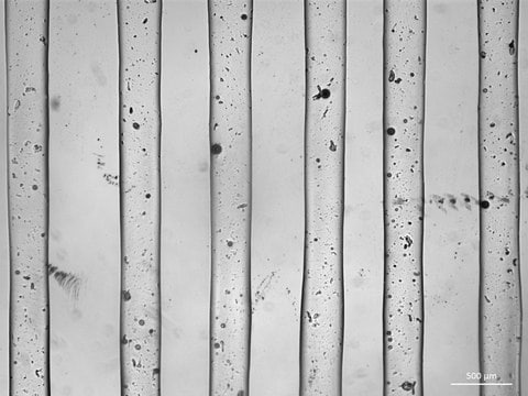
TissueFab® bioink Bone UV/365 nm, low endotoxin is a ready-to-use bioink which is formulated for high cell viability, osteoinduction and printing fidelity and is designed for extrusion-based 3D bioprinting and subsequent crosslinking with exposure to 405 nm visible light. GelMA-Bone bioinks can be used with most extrusion-based bioprinters, are biodegradable, and are compatible with human mesenchymal stem cells (hMSCs) and osteogenic cell types. TissueFab® bioink Bone UV/365 nm, low endotoxin enables the precise fabrication of osteogenic 3D cell models and tissue constructs for research in 3D cell biology, tissue engineering, in vitro tissue models, and regenerative medicine.
Legal Information
TISSUEFAB is a registered trademark of Merck KGaA, Darmstadt, Germany
Storage Class Code
10 - Combustible liquids
WGK
WGK 3
Choose from one of the most recent versions:
Certificates of Analysis (COA)
Lot/Batch Number
Sorry, we don't have COAs for this product available online at this time.
If you need assistance, please contact Customer Support.
Already Own This Product?
Find documentation for the products that you have recently purchased in the Document Library.
Nano hydroxyapatite particles promote osteogenesis in a three-dimensional bio-printing construct consisting of alginate/gelatin/hASCs.
Wang X F, et al.
Royal Society of Chemistry Advances, 6, 6832?42-6832?42 (2016)
Silke Wüst et al.
Acta biomaterialia, 10(2), 630-640 (2013-10-26)
Three-dimensional (3-D) bioprinting is the layer-by-layer deposition of biological material with the aim of achieving stable 3-D constructs for application in tissue engineering. It is a powerful tool for the spatially directed placement of multiple materials and/or cells within the
Mehdi Sadat-Shojai et al.
Materials science & engineering. C, Materials for biological applications, 49, 835-843 (2015-02-18)
The ability to encapsulate cells in three-dimensional (3D) protein-based hydrogels is potentially of benefit for tissue engineering and regenerative medicine. However, as a result of their poor mechanical strength, protein-based hydrogels have traditionally been considered for soft tissue engineering only.
Michal Bartnikowski et al.
Materials (Basel, Switzerland), 9(4) (2016-04-14)
The concept of biphasic or multi-layered compound scaffolds has been explored within numerous studies in the context of cartilage and osteochondral regeneration. To date, no system has been identified that stands out in terms of superior chondrogenesis, osteogenesis or the
Xi Chen et al.
International journal of nanomedicine, 11, 4707-4718 (2016-10-04)
Periodontitis is a chronic infectious disease and is the major cause of tooth loss and other oral health issues around the world. Periodontal tissue regeneration has therefore always been the ultimate goal of dentists and researchers. Existing fabrication methods mainly
Our team of scientists has experience in all areas of research including Life Science, Material Science, Chemical Synthesis, Chromatography, Analytical and many others.
Contact Technical Service
