Accéder au contenu
Merck
Toutes les photos(3)

Key Documents

M4276

Sigma-Aldrich

Monoclonal Anti-Myosin (Skeletal, Fast) antibody produced in mouse

clone MY-32, ascites fluid

Synonyme(s) :

Anti-Myosin Antibody

Se connecterpour consulter vos tarifs contractuels et ceux de votre entreprise/organisme


About This Item

Numéro MDL:
Code UNSPSC :
12352203
Nomenclature NACRES :
NA.41

Source biologique

mouse

Niveau de qualité

Conjugué

unconjugated

Forme d'anticorps

ascites fluid

Type de produit anticorps

primary antibodies

Clone

MY-32, monoclonal

Contient

15 mM sodium azide

Espèces réactives

rat, chicken, rabbit, mouse, human, bovine, guinea pig, feline

Technique(s)

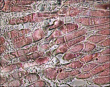
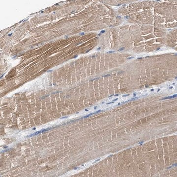
immunohistochemistry (formalin-fixed, paraffin-embedded sections): 1:400 using skeletal muscle tissue
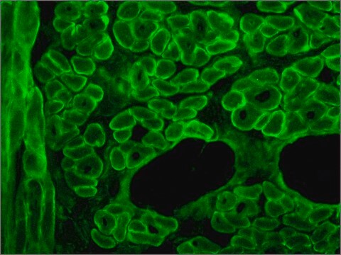
indirect immunofluorescence: 1:400
western blot: 1:1,000 using rabbit leg muscle extract

Isotype

IgG1

Numéro d'accès UniProt

Conditions d'expédition

dry ice

Température de stockage

−20°C

Modification post-traductionnelle de la cible

unmodified

Informations sur le gène

Vous recherchez des produits similaires ? Visite Guide de comparaison des produits

Description générale

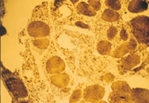
Localizes an epitope on the myosin heavy chain. Stains the fast (type II) and neonatal isomyosin molecules found in skeletal muscle, but does not stain cardiac muscle, smooth muscle or non-muscle myosin in cultured cells. Does react with human rhabdomyosarcomas.
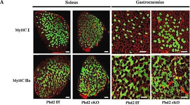
Monoclonal Anti-Skeletal Myosin (mouse IgG1 isotype) is derived from the hybridoma produced by the fusion of mouse myeloma cells and splenocytes from an immunized mouse. Myosin is a 480,000 dalton protein known to interact with actin in muscle and in non-muscle cells. It contains two identical heavy chains (200,000 daltons each) and four light chains (15,000-26,000 daltons). Myosin molecules consist of two major regions: tails (rods) and heads; they aggregate into filaments through the tail region and interact with actin and with ATP through the head region. Multiple forms of myosin heavy chains exist for each muscle type-skeletal, cardiac, smooth and non-muscle isomyosin forms exist in different types of skeletal muscle, depending on the physiological function of the muscle. These are designated at type I (slow twitch) and type II (fast-twitch). Type II fibers can be further subdivided in types IIA, IIB, and IIC.

Spécificité

Monoclonal Anti-Skeletal Myosin is specific for the myosin heavy chain. It does not stain human or animal cardiac or smooth muscle myosin or cells grown by tissue culture (nonmuscle myosin). It has been demonstrated on human skeletal muscle that the antibody stains the fast twitch (type II) isomyosin molecules. Monoclonal Anti-Skeletal Myosin antibody does react with human rhabdomyosarcomas.

Immunogène

rabbit muscle myosin.

Application

Monoclonal Anti-Myosin (Skeletal, Fast) antibody produced in mouse has been used in:
  • immunohistochemistry
  • immunostaining
  • western blotting at a dilution 1:1000 and 1:90000†
  • indirect immunofluorescence (dilution 1:400) of formalin-fixed, paraffin-embedded sections of human or animal skeletal muscle tissue preparation.
  • dot immunobinding on muscle extracts or purified myosin preparations
Monoclonal Anti-Skeletal Myosin may be used for staining of human, rabbit, rat, mouse, bovine, chicken and guinea pig skeletal myosin. Monoclonal Anti-Skeletal Myosin antibody to fast-twitch skeletal myosin may be used for detecting cross striated muscle differentiation in tumors. The antibody localizes an epitope on the myosin chain that is stable to the routine formalin-fixation and paraffin-embedding process.
The level of mysosin (fast) in serum samples from sportsmen with past injury was determined by western blot using monoclonal mouse anti-myosin (skeletal/fast) as the primary antibody at a dilution of 1:90000.

Clause de non-responsabilité

Unless otherwise stated in our catalog or other company documentation accompanying the product(s), our products are intended for research use only and are not to be used for any other purpose, which includes but is not limited to, unauthorized commercial uses, in vitro diagnostic uses, ex vivo or in vivo therapeutic uses or any type of consumption or application to humans or animals.

Vous ne trouvez pas le bon produit ?  

Essayez notre Outil de sélection de produits.

Code de la classe de stockage

10 - Combustible liquids

Classe de danger pour l'eau (WGK)

WGK 3

Point d'éclair (°F)

Not applicable

Point d'éclair (°C)

Not applicable


Certificats d'analyse (COA)

Recherchez un Certificats d'analyse (COA) en saisissant le numéro de lot du produit. Les numéros de lot figurent sur l'étiquette du produit après les mots "Lot" ou "Batch".

Déjà en possession de ce produit ?

Retrouvez la documentation relative aux produits que vous avez récemment achetés dans la Bibliothèque de documents.

Consulter la Bibliothèque de documents

Contractility and kinetics of human fetal and human adult skeletal muscle
Racca AW, et al.
The Journal of Physiology, 591(12), 3049-3061 (2013)
David Vaughan et al.
PloS one, 11(3), e0149046-e0149046 (2016-03-18)
A silencer region (I-allele) within intron 16 of the gene for the regulator of vascular perfusion, angiotensin-converting enzyme (ACE), is implicated in phenotypic variation of aerobic fitness and the development of type II diabetes. We hypothesised that the reportedly lower
Shanshan Wang et al.
Frontiers in cell and developmental biology, 8, 618617-618617 (2021-01-30)
Enhancer of zeste homolog 2 (EZH2) is the catalytic subunit of polycomb repressive complex 2 and contains a SET domain that catalyzes histone H3 trimethylation on lysine 27 (H3K27me3) to generate an epigenetic silencing mark. EZH2 interacts with transcription factors
An optimized histochemical method to assess skeletal muscle glycogen and lipid stores reveals two metabolically distinct populations of type I muscle fibers
Prats C, et al.
PLoS ONE, 8(10), e77774-e77774 (2013)
S Cefalù et al.
Cell cycle (Georgetown, Tex.), 14(6), 894-901 (2015-03-20)
p53 family members, p63 and p73, play a role in controlling early stage of myogenic differentiation. We demonstrated that TAp63gamma, unlike the other p53 family members, is markedly up-regulated during myogenic differentiation in murine C2C7 cell line. We also found

Notre équipe de scientifiques dispose d'une expérience dans tous les secteurs de la recherche, notamment en sciences de la vie, science des matériaux, synthèse chimique, chromatographie, analyse et dans de nombreux autres domaines..

Contacter notre Service technique