Accéder au contenu
Merck
Toutes les photos(4)

Principaux documents

HPA005539

Sigma-Aldrich

Anti-KANK1 antibody produced in rabbit

Prestige Antibodies® Powered by Atlas Antibodies, affinity isolated antibody, buffered aqueous glycerol solution

Synonyme(s) :

Anti-ANKRD15, Anti-Ankyrin repeat domain-containing protein 15 antibody produced in rabbit, Anti-Kidney ankyrin repeat-containing protein

Se connecterpour consulter vos tarifs contractuels et ceux de votre entreprise/organisme


About This Item

Code UNSPSC :
12352203
Numéro HPA (Human Protein Atlas):
Nomenclature NACRES :
NA.43

Source biologique

rabbit

Niveau de qualité

Conjugué

unconjugated

Forme d'anticorps

affinity isolated antibody

Type de produit anticorps

primary antibodies

Clone

polyclonal

Gamme de produits

Prestige Antibodies® Powered by Atlas Antibodies

Forme

buffered aqueous glycerol solution

Espèces réactives

human

Technique(s)

immunohistochemistry: 1:20- 1:50

Séquence immunogène

INVCGVRKRSYSAGNASQLEQLSRARRSGGELYIDYEEEEMETVEQSTQRIKEFRQLTADMQALEQKIQDSSCEASSELRENGECRSVAVGAEENMNDIVVYHRGSRSCKDAAVGTLVEMRNCGVSVTEAMLGVMTEADKEIE

Numéro d'accès UniProt

Conditions d'expédition

wet ice

Température de stockage

−20°C

Modification post-traductionnelle de la cible

unmodified

Informations sur le gène

human ... KANK1(23189)

Description générale

KN motif and ankyrin repeat domains 1 (KANK1) is an adaptor protein, which belongs to the KANK family of proteins. It contains ankyrin-repeat domain at its C-terminus, central coiled coil domains, and the KN motif at the N-terminal. KANK1 gene is located at the chromosomal position 9p24.3. This protein is found in both cytoplasm and nucleus, though it is predominantly found in the cytoplasm in multiple human kidney cell lines. Alternative splicing of this gene produces two isoforms, KANK-L and KANK-S, where KANK-L has an additional 158 amino acid at the N-terminal.

Immunogène

KN motif and ankyrin repeat domain-containing protein 1 recombinant protein epitope signature tag (PrEST)

Application

Anti-KANK1 antibody is suitable for immunoprecipitation and pull-down assay.
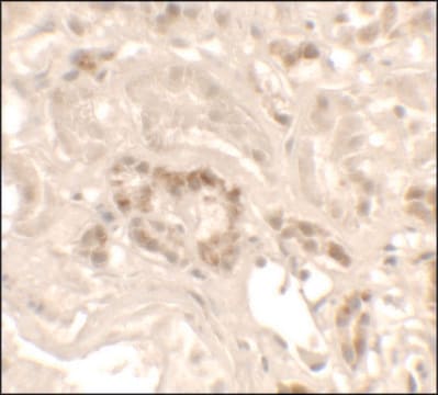
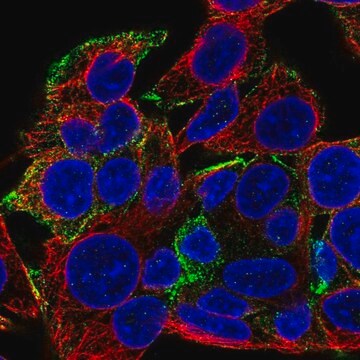
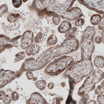
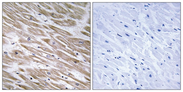
Anti-KANK1 antibody produced in rabbit, a Prestige Antibody, is developed and validated by the Human Protein Atlas (HPA) project . Each antibody is tested by immunohistochemistry against hundreds of normal and disease tissues. These images can be viewed on the Human Protein Atlas (HPA) site by clicking on the Image Gallery link. The antibodies are also tested using immunofluorescence and western blotting. To view these protocols and other useful information about Prestige Antibodies and the HPA, visit sigma.com/prestige.
Applications in which this antibody has been used successfully, and the associated peer-reviewed papers, are given below.
Western Blotting (1 paper)

Actions biochimiques/physiologiques

KANK1 (KN motif and ankyrin repeat domains 1) protein regulates the formation of cytoskeleton by mediating actin polymerization. Akt phosphorylates KANK1, which in turn binds to 14-3-3 protein. The activation of Akt is dependent upon epidermal growth factor (EGF) and insulin, which control cell migration, apoptosis, cell cycle and growth etc. Through Akt signaling, KANK1 suppresses cell migration and actin stress fiber formation by inhibiting RhoA. It prevents the formation of lamellipodia in fibroblasts via its interaction with IRSp53. It also negatively regulates integrin dependent cell spreading. KANK1 acts as a nucleo-cytoplasmic shuttle for β-catenin protein, where β-catenin is transported to nucleus to induce transcription dependent on β-catenin. KANK1 also acts as a tumor suppressor gene, and its down-regulation or inactivation is associated with kidney cancer, prostate cancer, lung cancer, bladder cancer, and breast cancer.

Caractéristiques et avantages

Prestige Antibodies® are highly characterized and extensively validated antibodies with the added benefit of all available characterization data for each target being accessible via the Human Protein Atlas portal linked just below the product name at the top of this page. The uniqueness and low cross-reactivity of the Prestige Antibodies® to other proteins are due to a thorough selection of antigen regions, affinity purification, and stringent selection. Prestige antigen controls are available for every corresponding Prestige Antibody and can be found in the linkage section.

Every Prestige Antibody is tested in the following ways:
  • IHC tissue array of 44 normal human tissues and 20 of the most common cancer type tissues.
  • Protein array of 364 human recombinant protein fragments.

Liaison

Corresponding Antigen APREST85227

Forme physique

Solution in phosphate-buffered saline, pH 7.2, containing 40% glycerol and 0.02% sodium azide

Informations légales

Prestige Antibodies is a registered trademark of Merck KGaA, Darmstadt, Germany

Clause de non-responsabilité

Unless otherwise stated in our catalog or other company documentation accompanying the product(s), our products are intended for research use only and are not to be used for any other purpose, which includes but is not limited to, unauthorized commercial uses, in vitro diagnostic uses, ex vivo or in vivo therapeutic uses or any type of consumption or application to humans or animals.

Vous ne trouvez pas le bon produit ?  

Essayez notre Outil de sélection de produits.

Code de la classe de stockage

10 - Combustible liquids

Classe de danger pour l'eau (WGK)

WGK 1

Point d'éclair (°F)

Not applicable

Point d'éclair (°C)

Not applicable

Équipement de protection individuelle

Eyeshields, Gloves, multi-purpose combination respirator cartridge (US)


Certificats d'analyse (COA)

Recherchez un Certificats d'analyse (COA) en saisissant le numéro de lot du produit. Les numéros de lot figurent sur l'étiquette du produit après les mots "Lot" ou "Batch".

Déjà en possession de ce produit ?

Retrouvez la documentation relative aux produits que vous avez récemment achetés dans la Bibliothèque de documents.

Consulter la Bibliothèque de documents

Xiaohang Guo et al.
International journal of oncology, 44(3), 797-804 (2014-01-09)
The Kank1 gene is one of the important members of the Kank gene family. As an important adaptor protein, Kank1 plays a significant role in the genesis and development of many malignant tumors. It was recently discovered that the Kank1
Rena J Vanzo et al.
European journal of medical genetics, 56(5), 256-259 (2013-03-05)
Deletion of the KANK1 gene (also called ANKRD15), located at chromosome position 9p24.3, has been associated with neurodevelopmental disease including congenital cerebral palsy, hypotonia, quadriplegia, and intellectual disability in a four-generation family. The inheritance pattern in this family was suggested
Yong Wang et al.
Journal of cell science, 119(Pt 19), 4002-4010 (2006-09-14)
The human Kank protein has a role in controlling the formation of the cytoskeleton by regulating actin polymerization. Besides the cytoplasmic localization as reported before, we observed the nuclear localization of Kank in OS-RC-2 cells. To uncover the mechanism behind
Yong Wang et al.
Biochemical and biophysical research communications, 330(4), 1247-1253 (2005-04-13)
The human Kank gene encodes an ankyrin repeat domain-containing protein which regulates actin polymerization. There are at least two types of Kank protein depending on cell type, likely due to differences in transcription. Here, to examine the transcriptional initiation and
N Kakinuma et al.
Cellular and molecular life sciences : CMLS, 66(16), 2651-2659 (2009-06-26)
The Kank family of proteins, Kank1-Kank4, are characterized by their unique structure, coiled-coil motifs in the N-terminal region, and ankyrin-repeats in the C-terminal region, with an additional motif, the KN motif, at the N-terminus. Kank1 was obtained by positional cloning

Notre équipe de scientifiques dispose d'une expérience dans tous les secteurs de la recherche, notamment en sciences de la vie, science des matériaux, synthèse chimique, chromatographie, analyse et dans de nombreux autres domaines..

Contacter notre Service technique