Accéder au contenu
Merck
Toutes les photos(1)

Key Documents

A2080

Sigma-Aldrich

Monoclonal Anti-Alkaline Phosphatase, Human Placental−Agarose antibody produced in mouse

clone 8B6, purified immunoglobulin, PBS suspension

Synonyme(s) :

Monoclonal Anti-Alkaline Phosphatase, Human Placental, Anti-Phosphatase, Alkaline, Human Placental, PLAP

Se connecterpour consulter vos tarifs contractuels et ceux de votre entreprise/organisme


About This Item

Numéro MDL:
Code UNSPSC :
12352203
Nomenclature NACRES :
NA.46

Source biologique

mouse

Niveau de qualité

Conjugué

agarose conjugate

Forme d'anticorps

purified immunoglobulin

Type de produit anticorps

primary antibodies

Clone

8B6, monoclonal

Forme

PBS suspension

Espèces réactives

human

Ampleur du marquage

2 mg antibody per mL bed volume

Technique(s)

immunoprecipitation (IP): suitable

Isotype

IgG2a

Numéro d'accès UniProt

Conditions d'expédition

wet ice

Température de stockage

2-8°C

Modification post-traductionnelle de la cible

unmodified

Informations sur le gène

human ... ALPP(250)

Vous recherchez des produits similaires ? Visite Guide de comparaison des produits

Description générale

In SDS gels, the product reacts with both Regan and Nagao isozymes of human placental alkaline phosphatase (hPLAP, 130 kDa, 67/130 kDa). By RIA, the antibody binds to hPLAP with an affinity constant of 5 × 10⁹ LM-1. It does not react with PLAP-like enzymes.
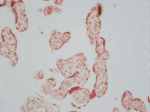
Monoclonal Anti-Human Placental Alkaline Phosphatase (mouse IgG2a isotype) is derived from the 8B6 hybridoma produced by the fusion of mouse myeloma cells and splenocytes from immunized BALB/c mice. Alkaline Phosphatase (AP) is a broad and general term associated with non-specific phosphomonoesterases, with optimal activity at alkaline pH. Human placental alkaline phosphatase (EC 3.1.3.1; hPLAP), an isoenzyme of the AP group of enzymes, is ordinarily synthesized in the placental syncytiotrophoblast, becoming detectable in maternal circulation after the twelfth week of pregnancy. Small amounts of hPLAP are also found in the endocervix, fallopian tubes and lung. Very small amounts of heat-stable AP resembling hPLAP (hPLAP-like AP) are expressed in the testis, thymus and in rare colon epithelial cells.

Immunogène

Human epidermoid carcinoma cell line expressing placental alkaline phosphatase.

Application

Anti-Alkaline phosphatase human placental antibody coupled to agarose was packed in a column and used to purify alkaline-phosphatase conjugated FGF receptor out of conditioned media from COS-7 cells. Column was washed with 1M NaCl.
Cultured pancreatic islet cells were fixed with cold 4% paraformaldhyde at room temperature for 10 minutes and incubated with mouse monoclonal anti-Alkaline Phosphatase, Human Placental antibody at 1:500. Mouse monoclonal anti-Alkaline Phosphatase, Human Placental antibody was used to detect a unique molecular probe that consisted of the FGFR extracellular domain expressed as a chimera with human PLAP.
This immunoadsorbant may be used to purify fusion proteins produced using vectors expressing the hPLAP gene. Suitable for affinity purification and immunoprecipitation procedures of human placental alkaline phosphatase either natural or recombinant.It is alsu used in western blotting.

Actions biochimiques/physiologiques

Alkaline phosphatases are non-specific phosphomonoesterases that function optimally at alkaline pH. Placental alkaline phosphatase differs from the alkaline phosphatases of bone, liver and kidney, in the hydrolysis rate of various phosphate-based substrates, and also in its degree of inhibition by L-phenylalanine and L-homoarginine or levamisole. Human Placental Alkaline Phosphatase (hPLAP) has been studied as a potential tumor marker.
The human placental alkaline phosphatase (hPLAP and hPLAP-like AP) is present in the sera and tumor tissue of patients suffering from various types of cancer. Despite the continued interest in hPLAP as a potential tumor marker, it has not been widely used in the routine clinical laboratory, mainly because of the low overall specificity attained by methodologies such as heat-inactivation and sensitivity to L-phenylalanine.

Forme physique

Suspension in 0.1 M phosphate buffered saline, pH 7.4, containing 15 mM sodium azide.

Notes préparatoires

Prepared using purified antibody coupled to cyanogen bromide-activated agarose.

Clause de non-responsabilité

Unless otherwise stated in our catalog or other company documentation accompanying the product(s), our products are intended for research use only and are not to be used for any other purpose, which includes but is not limited to, unauthorized commercial uses, in vitro diagnostic uses, ex vivo or in vivo therapeutic uses or any type of consumption or application to humans or animals.

Vous ne trouvez pas le bon produit ?  

Essayez notre Outil de sélection de produits.

Code de la classe de stockage

10 - Combustible liquids

Classe de danger pour l'eau (WGK)

WGK 3

Point d'éclair (°F)

Not applicable

Point d'éclair (°C)

Not applicable


Certificats d'analyse (COA)

Recherchez un Certificats d'analyse (COA) en saisissant le numéro de lot du produit. Les numéros de lot figurent sur l'étiquette du produit après les mots "Lot" ou "Batch".

Déjà en possession de ce produit ?

Retrouvez la documentation relative aux produits que vous avez récemment achetés dans la Bibliothèque de documents.

Consulter la Bibliothèque de documents

Songshan Ye et al.
Molecular medicine reports, 13(6), 4593-4598 (2016-04-16)
Paeoniflorin, the major component of Paeonia lactiflora pall, has previously been reported to prevent thrombosis. Plasminogen activator urokinase (uPA) is a serine protease that markedly facilitates normal thrombosis resolution. Paeoniflorin and uPA have been linked to the mitogen‑activated protein kinase (MAPK)
Extremely high maternal alkaline phosphatase serum concentration with syncytiotrophoblastic origin
Boronkai A, et al.
Journal of Clinical Pathology, 58(1), 72-76 (2005)
Adam W Barb et al.
Protein science : a publication of the Protein Society, 21(10), 1456-1466 (2012-08-02)
Paramagnetic lanthanide ions when bound to proteins offer great potential for structural investigations that utilize solution nuclear magnetic resonance spectroscopy, magnetic resonance imaging, or optical microscopy. However, many proteins do not have native metal ion binding sites and engineering a
Alkaline phosphatases
Millan JL
Purinergic Signaling, 2(2), 335-335 (2006)
Xinghua Hou et al.
The Journal of biological chemistry, 287(1), 474-483 (2011-11-15)
NOTCH signaling induced by Delta1 (DLL1) and Jagged1 (JAG1) NOTCH ligands is modulated by the β3N-acetylglucosaminyl transferase Fringe. LFNG (Lunatic Fringe) and MFNG (Manic Fringe) transfer N-acetylglucosamine (GlcNAc) to O-fucose attached to EGF-like repeats of NOTCH receptors. In co-culture NOTCH

Notre équipe de scientifiques dispose d'une expérience dans tous les secteurs de la recherche, notamment en sciences de la vie, science des matériaux, synthèse chimique, chromatographie, analyse et dans de nombreux autres domaines..

Contacter notre Service technique