Accéder au contenu
Merck
Toutes les photos(2)

Key Documents

MABS2029

Sigma-Aldrich

Anti-YAP Antibody, clone 8G5

clone 8G5, from rat

Synonyme(s) :

Transcriptional coactivator YAP1, Yes-associated protein 1, Protein yorkie homolog, Yes-associated protein YAP65 homolog

Se connecterpour consulter vos tarifs contractuels et ceux de votre entreprise/organisme


About This Item

Code UNSPSC :
12352203
eCl@ss :
32160702
Nomenclature NACRES :
NA.41

Source biologique

rat

Forme d'anticorps

purified immunoglobulin

Type de produit anticorps

primary antibodies

Clone

8G5, monoclonal

Espèces réactives

mouse, human

Conditionnement

antibody small pack of 25 μg

Technique(s)

immunocytochemistry: suitable
immunofluorescence: suitable
immunohistochemistry: suitable (paraffin)
immunoprecipitation (IP): suitable
western blot: suitable

Isotype

IgG2aκ

Numéro d'accès NCBI

Numéro d'accès UniProt

Modification post-traductionnelle de la cible

unmodified

Informations sur le gène

human ... YAP1(10413)

Description générale

Transcriptional coactivator YAP1 (UniProt: P46937; also known as Yes-associated protein 1, Protein yorkie homolog, Yes-associated protein YAP65 homolog) is encoded by the YAP1 (also known as YAP65) gene (Gene ID: 10413) in human. YAP1 is a transcriptional regulator that can act both as a coactivator and a corepressor and is the critical downstream regulatory target in the Hippo signaling pathway. It also plays a key role in tissue tension and 3D tissue shape by regulating cortical actomyosin network formation. YAP1 contains two WW domains (aa 171-204 and 230-263) and a transactivation domain (aa 291-504). It also has two coiled coil regions (aa 86-100 and 298-359) and the first coiled-coil region is reported to mediate most of the interaction with TEAD transcription factors. The presence of TEAD transcription factors is required for YAP1 to stimulate gene expression, cell growth, anchorage-independent growth, and epithelial mesenchymal transition (EMT) induction. YAP1 phosphorylation status and cell density can regulate its subcellular localization. Its phosphorylation by LATS1 and LATS2 leads to its cytoplasmic translocation and inactivation. YAP1 can also be phosphorylated by ABL1, which leads to its stabilization, increased interaction with TP73, and recruitment onto proapoptotic genes in response to DNA damage. Nine isoforms of YAP1 have been described that are produced by alternative splicing. Mutations in YAP1 gene have been linked to ocular coloboma that is characterized by uveal colobomata, microphthalmia, cataract and cleft lip/palate. Higher expression of YAP1 has been reported in some liver and prostate cancers.

Spécificité

Clone 8G5 is a rat monoclonal antibody that detects Transcriptional coactivator YAP1. It targets an epitope with in 12 amino acids from the C-terminal region.

Immunogène

KLH-conjugated linear peptide corresponding to 12 amino acids from the C-terminal region of human Transcriptional coactivator YAP1.

Application

Anti-YAP, clone 8G5, Cat. No. MABS2029, is a rat monoclonal antibody that detects Transcriptional coactivator YAP1 and has been tested or use in Immunocytochemistry, Immunofluorescence, Immunohistochemistry (Paraffin), Immunoprecipitation, and Western Blotting.
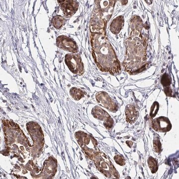
Immunohistochemistry (Paraffin) Analysis: A 1:50 dilution from a representative lot detected YAP in mouse colon tissue sections.

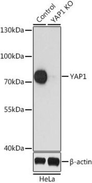
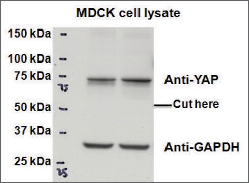
Western Blotting Analysis: A representative lot detected YAP in Western Blotting applications (Miyamura, N.,et. al. (2017). Nat Commun. 8:16017; Matsudaira, T., et. al. (2017). Nat Commun. 8(1):1246; Hata, S., et. al. (2012). J Biol Chem. 287(26):22089-98; Maruyama, J., et. al. (2017). Mol Cancer Res. 16(2):197-211).

Immunoprecipitation Analysis: A representative lot detected YAP in Immunoprecipitation applications (Hata, S., et. al. (2012). J Biol Chem. 287(26):22089-98).

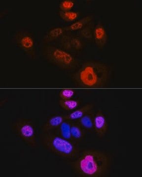
Immunofluorescence Analysis: A representative lot detected YAP in Immunofluorescence applications (Miyamura, N.,et. al. (2017). Nat Commun. 8:16017).

Immunocytochemistry Analysis: A representative lot detected YAP in Immunocytochemistry applications (Matsudaira, T., et. al. (2017). Nat Commun. 8(1):1246; Hata, S., et. al. (2012). J Biol Chem. 287(26):22089-98).

Qualité

Evaluated by Immunohistochemistry (Paraffin) in human placenta tissue sections.

Immunohistochemistry (Paraffin) Analysis: A 1:50 dilution of this antibody detected YAP in human placenta tissue sections.

Description de la cible

54.46 kDa calculated.

Forme physique

Format: Purified

Autres remarques

Concentration: Please refer to lot specific datasheet.

Vous ne trouvez pas le bon produit ?  

Essayez notre Outil de sélection de produits.


Certificats d'analyse (COA)

Recherchez un Certificats d'analyse (COA) en saisissant le numéro de lot du produit. Les numéros de lot figurent sur l'étiquette du produit après les mots "Lot" ou "Batch".

Déjà en possession de ce produit ?

Retrouvez la documentation relative aux produits que vous avez récemment achetés dans la Bibliothèque de documents.

Consulter la Bibliothèque de documents

Sakurako Kobayashi et al.
Inflammation and regeneration, 42(1), 49-49 (2022-11-30)
The emerging concepts of fetal-like reprogramming following tissue injury have been well recognized as an important cue for resolving regenerative mechanisms of intestinal epithelium during inflammation. We previously revealed that the remodeling of mesenchyme with collagen fibril induces YAP/TAZ-dependent fate

Notre équipe de scientifiques dispose d'une expérience dans tous les secteurs de la recherche, notamment en sciences de la vie, science des matériaux, synthèse chimique, chromatographie, analyse et dans de nombreux autres domaines..

Contacter notre Service technique