Accéder au contenu
Merck
Toutes les photos(2)

Key Documents

MABN153

Sigma-Aldrich

Anti-HuC/HuD Antibody, clone 15A7.1

clone 15A7.1, from mouse

Synonyme(s) :

ELAV-like protein 3, Hu-antigen C, HuC, Paraneoplastic cerebellar degeneration-associated antigen, Paraneoplastic limbic encephalitis antigen 21, ELAV-like protein 4, Hu-antigen D, HuD, Paraneoplastic encephalomyelitis antigen HuD

Se connecterpour consulter vos tarifs contractuels et ceux de votre entreprise/organisme


About This Item

Code UNSPSC :
12352203
eCl@ss :
32160702
Nomenclature NACRES :
NA.41

Source biologique

mouse

Niveau de qualité

Forme d'anticorps

purified antibody

Type de produit anticorps

primary antibodies

Clone

15A7.1, monoclonal

Espèces réactives

human, rat, mouse

Technique(s)

immunohistochemistry: suitable (paraffin)
western blot: suitable

Isotype

IgG2aκ

Numéro d'accès NCBI

Numéro d'accès UniProt

Conditions d'expédition

wet ice

Modification post-traductionnelle de la cible

unmodified

Informations sur le gène

human ... ELAVL4(1996)

Description générale

Hu proteins are mammalian embryonic lethal abnormal visual system (ELAV)-like neuronal RNA-binding proteins that contain three RNA recognition motifs. RNA binding protein HuC promotes neuronal development in both the central and the peripheral nervous systems. HuC antibodies are used as markers for developing neurons. HuD may play a role in neuron-specific RNA processing, and be involved in maintaining the proliferative state of cells.

Immunogène

KLH-conjugated linear peptide corresponding to human HuC/HuD.

Application

&
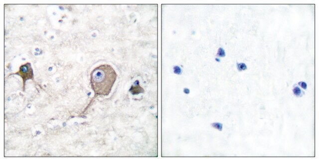
Analyse par immunohistochimie : A 1:2,000 dilution from a representative lot detected HuC/HuD in human brain and rat hippocampus tissues.
Domaine de recherche
Neurosciences
Sous-domaine de recherche
Signalisation du développement

Qualité

Produit évalué par western blotting sur du lysat de tissu cérébral humain.

Analyse par western blotting : µµ

Description de la cible

Poids réel (observé) : env. 40 kDa. An uncharacterized band may appear at ~45 kDa in some lysates.

Forme physique

Anticorps IgG2aκ monoclonal de souris purifié, dans un tampon contenant 0,1 M de Tris-glycine, pH 7,4, 150 mM de NaCl et 0,05 % d'azoture de sodium.
Format : Produit purifié
Produit purifié sur protéine G

Stockage et stabilité

Stable entre 2 et 8 °C pendant 1 an à compter de la date de réception.

Remarque sur l'analyse

Témoin
Lysat de tissu cérébral humain

Autres remarques

Concentration : pour connaître la concentration spécifique du lot, voir le certificat d'analyse.

Clause de non-responsabilité

Sauf indication contraire dans notre catalogue ou toute autre documentation associée au(x) produit(s), nos produits sont uniquement destinés à la recherche et ne sauraient être utilisés à d'autres fins, ce qui inclut, sans s'y limiter, les utilisations commerciales non autorisées, les utilisations diagnostiques in vitro, les utilisations thérapeutiques ex vivo ou in vivo, ou tout type de consommation ou d'application chez l'être humain ou chez l'animal.

Vous ne trouvez pas le bon produit ?  

Essayez notre Outil de sélection de produits.

Code de la classe de stockage

12 - Non Combustible Liquids

Classe de danger pour l'eau (WGK)

WGK 1

Point d'éclair (°F)

Not applicable

Point d'éclair (°C)

Not applicable


Certificats d'analyse (COA)

Recherchez un Certificats d'analyse (COA) en saisissant le numéro de lot du produit. Les numéros de lot figurent sur l'étiquette du produit après les mots "Lot" ou "Batch".

Déjà en possession de ce produit ?

Retrouvez la documentation relative aux produits que vous avez récemment achetés dans la Bibliothèque de documents.

Consulter la Bibliothèque de documents

Joslyn Lay et al.
American journal of physiology. Gastrointestinal and liver physiology, 311(2), G252-G266 (2016-07-02)
The μ-opioid receptor (MOR) is a major regulator of gastrointestinal motility and secretion and mediates opiate-induced bowel dysfunction. Although MOR is of physiological and therapeutic importance to gut function, the cellular and subcellular distribution and regulation of MOR within the
Christine Marques et al.
Acta neuropathologica, 147(1), 56-56 (2024-03-13)
The stimulator of interferon genes (STING) pathway has been implicated in neurodegenerative diseases, including Parkinson's disease and amyotrophic lateral sclerosis (ALS). While prior studies have focused on STING within immune cells, little is known about STING within neurons. Here, we
Karolína Liška et al.
Neuroendocrinology, 112(4), 384-398 (2021-06-11)
Circadian clocks in the hippocampus (HPC) align memory processing with appropriate time of day. Our study was aimed at ascertaining the specificity of glycogen synthase kinase 3-beta (GSK3β)- and glucocorticoid (GC)-dependent pathways in the entrainment of clocks in individual HPC
Yaxing Gui et al.
Brain pathology (Zurich, Switzerland), 30(1), 151-164 (2019-07-06)
The 18 kDa translocator protein (TSPO) is a widely used target for microglial PET imaging radioligands, but its expression in post-mortem normal and diseased human brain is not well described. We aimed at characterizing the TSPO expression in human control
Clara Muñoz-Castro et al.
Methods in molecular biology (Clifton, N.J.), 2593, 283-305 (2022-12-14)
There is a growing interest in expanding the multiplexing capability of immunohistochemistry to achieve a deeper phenotyping of various cell types in health and disease. Here, we describe a protocol of cyclic multiplex fluorescent immunohistochemistry that enables the labeling of

Notre équipe de scientifiques dispose d'une expérience dans tous les secteurs de la recherche, notamment en sciences de la vie, science des matériaux, synthèse chimique, chromatographie, analyse et dans de nombreux autres domaines..

Contacter notre Service technique